Medical care in space has long faced a quiet but serious limitation. For more than 40 years, astronauts relied on ultrasound as their only imaging tool. While useful, it cannot answer every question when something goes wrong.
Now, a new study shows a major step forward. Crew members aboard a commercial spaceflight have captured the first diagnostic X-rays in orbit. The results suggest that a simple, portable device could transform how health and safety are managed in space.
Space travel places the human body under strain. Bones weaken, fluids shift, and injuries become harder to diagnose. As missions grow longer, risks increase.
Ultrasound has served as the main imaging tool because it is compact and safe. Still, it has limits. It depends heavily on operator skill and cannot always detect fractures or lung problems.
Dr. Sheyna Gifford, lead researcher and assistant professor of aerospace medicine at Mayo Clinic, explained the challenge. “It’s been a dream for aerospace medicine to have more than one imaging modality for diagnosing illnesses and injuries in space,” she said. “X-rays are fast, easy and diagnostically valuable.”
Until recently, many believed X-rays would not work in orbit. Traditional machines are large, sensitive to motion, and produce radiation. In microgravity, where everything floats, clear imaging seemed unlikely.
Advances in portable radiography changed that assumption. Small, battery-powered X-ray systems are now used in sports arenas, remote clinics, and emergency settings.
Researchers saw a chance to test these devices in space. Earlier experiments during parabolic flights showed promise. Crew members successfully captured a hand X-ray in brief periods of weightlessness.
Building on that success, the team partnered with SpaceX for a full orbital test. The mission, known as Fram2, lasted three and a half days in a polar orbit between 425 and 450 kilometers above Earth.
Before launch, three crew members completed just four hours of training. They learned how to operate the system and position subjects for imaging.
The goal was simple. Could people without medical backgrounds capture useful X-rays in space?
The system itself was compact and wireless. It included a digital X-ray generator and a flat panel detector. Engineers tested it against vibration, extreme temperatures, and vacuum conditions before flight.
During the mission, crew members captured images without real-time help from Earth. They followed written and visual instructions.
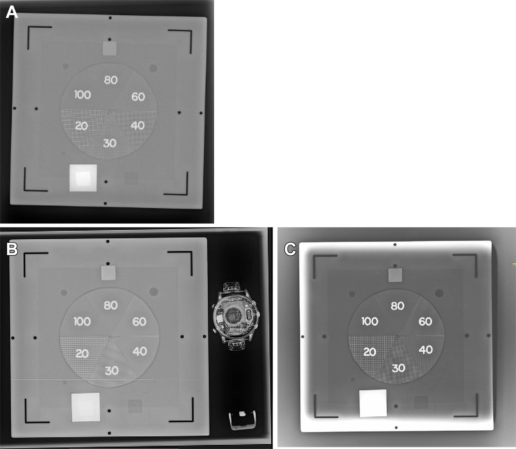
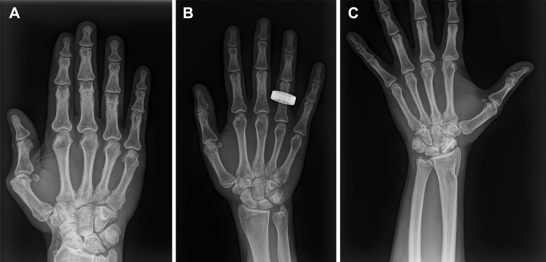
They imaged both the human body and objects inside the spacecraft. These included a hand, forearm, chest, abdomen, pelvis, and a smartwatch.
Images were transmitted instantly to onboard computers for review. After the mission, additional images were taken on Earth for comparison.
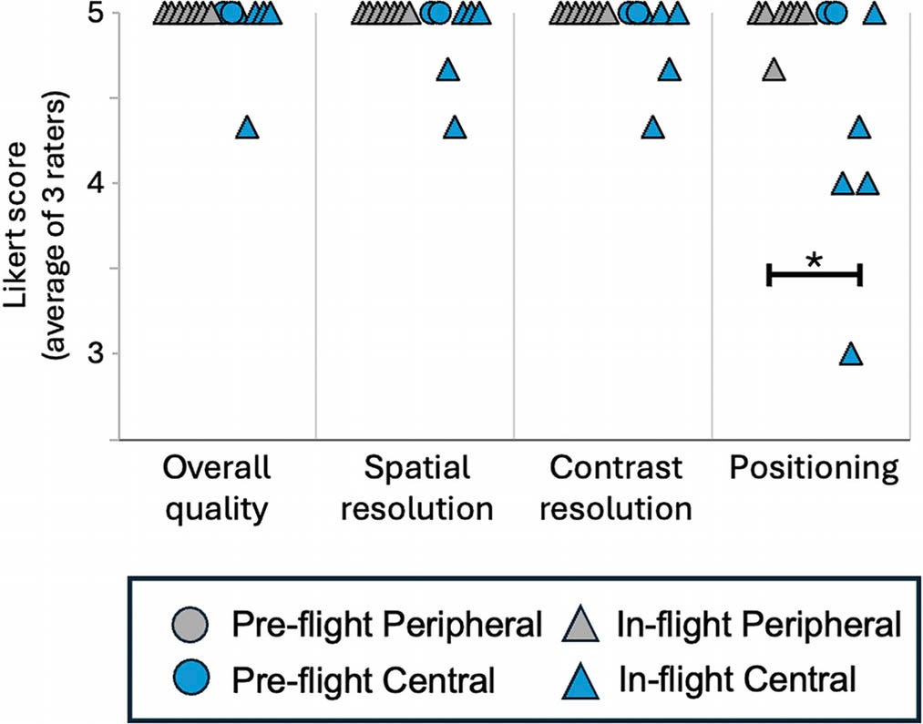
Radiologists later evaluated all images. They assessed clarity, detail, contrast, and positioning.

The results were strong. Image quality in space matched that of images taken before the flight. There were no meaningful differences in resolution or contrast.
Every image met diagnostic standards. Most were rated good to excellent.
Dr. Gifford emphasized the achievement. “Acquiring a diagnostically useful X-rays in space is something that anyone can do,” she said. “Three very talented nonmedical people with four hours of training in one of the harshest environments did it right and did it well.”
One difficulty did emerge. Positioning the body for imaging proved harder in microgravity.
Without gravity, both the patient and equipment tend to drift. This made it more difficult to align the X-ray source and detector, especially for larger body areas.
Images of the chest, abdomen, and pelvis showed slightly lower positioning scores than those taken on Earth. However, the images were still clear enough for diagnosis.
Smaller areas, such as the hand and forearm, showed no positioning issues.
![Charts show crewmember survey results regarding the operational use of the x-ray system (n = 3, 2 days after return [R+2]).](https://www.thebrighterside.news/uploads/2026/07/space-xray.jpg)
Crew members suggested simple improvements. They recommended clamps, mounts, and better ways to secure the equipment.
The study also explored another important use. X-rays can inspect equipment without taking it apart.
This method, known as nondestructive testing, allows engineers to look inside tools, electronics, and spacesuits.
Images of a smartwatch revealed fine internal details. The system captured structures at a very small scale, showing strong resolution.
This capability could prove vital on long missions. Detecting hidden damage early could prevent serious failures.
Dr. Gifford highlighted this broader impact. “A spaceflight-ready radiography system would have profound implications not only for crew health but also for mission-critical nonmedical tasks,” she said.
Radiation exposure was carefully monitored during the mission. Total exposure over the three and a half days reached about 1.5 millisieverts.
This includes both natural space radiation and the X-rays used in the study. The additional exposure from imaging was similar to standard medical scans on Earth.
Other crew members received minimal scattered radiation. These levels were very low and considered safe.
After the spacecraft returned to Earth, researchers examined the equipment. The X-ray generator showed minor external damage from landing. Internal components remained intact.
The detector also performed well. Image quality stayed consistent, with no significant loss of function.
This durability is critical. Space equipment must survive harsh conditions, including launch and reentry.
The success of this experiment marks a turning point. Astronauts may soon have access to faster and more reliable diagnostics.
X-ray imaging can help detect fractures, lung conditions, and internal injuries. It can also track bone density loss over time.
As missions extend to the Moon and Mars, these capabilities will become essential. Crews will need to diagnose and manage health issues without immediate help from Earth.
This study opens new possibilities for both space and Earth. In orbit, portable X-ray systems could improve medical care and equipment monitoring. They provide fast, clear insights that were not previously available.
For future missions, these systems could support longer journeys and reduce risks. Astronauts may rely on them to diagnose injuries, guide treatment, and maintain critical hardware.
On Earth, the same technology could benefit remote and underserved communities. Portable systems that require little training could bring medical imaging to places without hospitals.
The research also highlights the value of simple, adaptable tools. By using commercially available equipment, the team showed that advanced care does not always require complex systems.
The first X-rays taken in orbit mark more than a technical milestone. They represent a shift in how humans prepare for life beyond Earth.
With minimal training, small devices, and careful planning, crew members achieved something once thought impossible.
The images are clear. The system works. The path forward is now visible.
As space exploration continues, tools like these will help ensure that crews remain safe, informed, and ready for whatever challenges lie ahead.
Research findings are available online in the journal Radiology.
The original story “First x-rays captured in space mark breakthrough for astronaut health” is published in The Brighter Side of News.
Like these kind of feel good stories? Get The Brighter Side of News’ newsletter.
The post First x-rays captured in space mark breakthrough for astronaut health appeared first on The Brighter Side of News.

