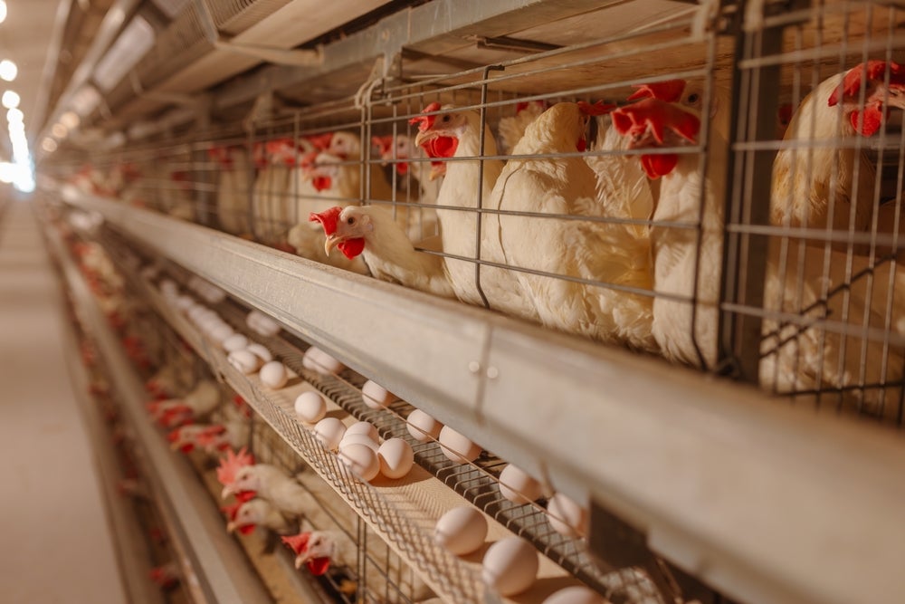
Eggs may look simple from the outside, but each one can hold valuable information about life, health and future production. For hatcheries, that hidden information matters. A single egg may contain a healthy embryo, a dead embryo, an unfertilized yolk or a chick that will later be culled after hatching.
Researchers at the University of Illinois Urbana-Champaign are working to make that hidden world visible without cracking the shell. Their studies use near infrared imaging, hyperspectral imaging and machine learning to evaluate eggs quickly and safely. The goal is to help hatcheries improve production while reducing waste, disease risk and animal welfare concerns.
The work could change how the poultry industry handles millions of eggs. Instead of relying on slow, labor-heavy checks, hatcheries may one day scan eggs automatically. A computer model could then identify which eggs are viable, which embryos have died and even which eggs contain male or female embryos.
Traditional egg testing often requires breaking eggs or relying on visual inspection. Hatcheries commonly use candling, which shines bright light through an egg to check development. While useful, candling takes time, depends on worker judgment and can miss early problems.
The Illinois team tested whether imaging tools could do more. Hyperspectral imaging, or HSI, captures hundreds of light bands across the visible and near infrared spectrum. These bands reveal subtle chemical and biological clues hidden inside the egg.
Near infrared spectroscopy, or NIR, captures fewer bands but costs less. It can still detect useful information about shell strength, shell thickness and yolk ratio. Both methods allow researchers to study eggs without damaging them.
“With NIR and HSI, we do not need to destroy the eggs. We just need to scan them and the machine learning model will determine the desired parameter,” said Mohammed Kamruzzaman, assistant professor in Agricultural and Biological Engineering.
In one recent study, researchers focused on chick embryo mortality. Hatcheries can lose more than 10% of embryos, which affects profit, efficiency and animal welfare. Dead embryos can also harbor bacteria, creating biosecurity concerns.
“If we can detect and remove them early in the incubation period, we can avoid biosecurity issues,” said lead author Md. Wadud Ahmed.
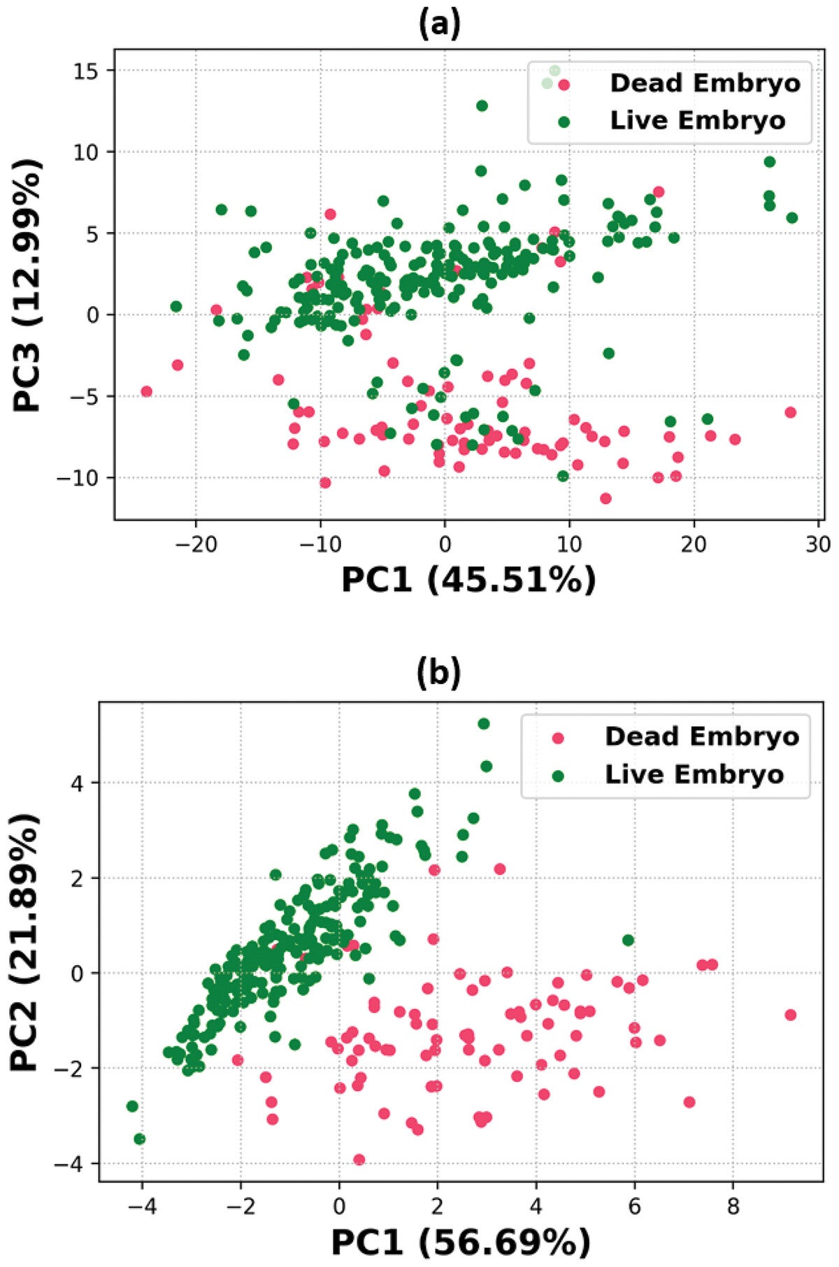
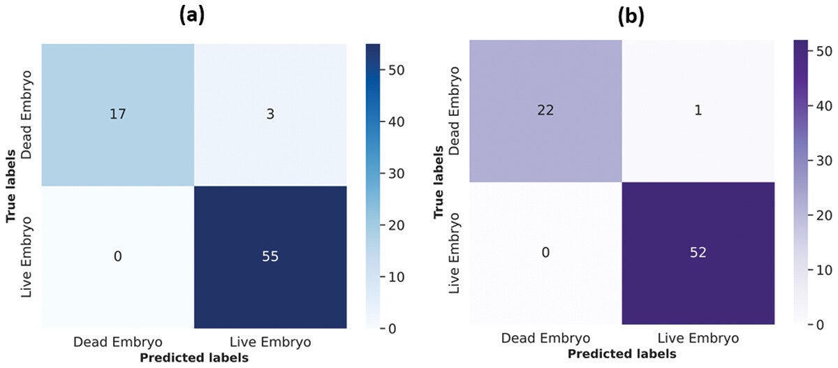
The team collected 300 chicken eggs from the university poultry farm and placed them in a commercial incubator. They captured hyperspectral images before incubation and again after four days. After incubation, they identified which embryos were alive and which had died.
The researchers then trained machine learning models to read spectral patterns linked to embryo survival. By day four, the best model reached up to 97% accuracy. A feature-selected model later reached 98.7% test accuracy.
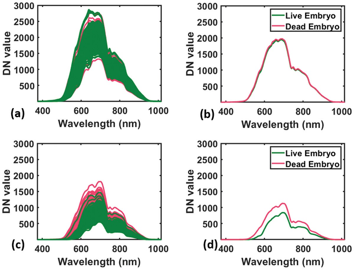
The images showed clear biological differences. Live embryos absorbed more light in regions linked to blood development, water balance and energy use. Dead embryos appeared brighter across many wavelengths because they lacked normal blood vessel formation and metabolism.
The strongest signals appeared between 500 and 900 nanometers. Some bands reflected carotenoids in the yolk, which support embryo growth. Others reflected hemoglobin, the oxygen-carrying molecule tied to developing blood vessels.
Wavelengths near 750 and 773 nanometers pointed to water and protein changes. A band near 837 nanometers reflected lipids, which embryos use for energy. Together, these markers helped the models separate viable embryos from failing ones.
The team also used explainable artificial intelligence to understand model decisions. One key wavelength, 709 nanometers, related to hydration and tissue development. Another, 545 nanometers, reflected oxygen-rich blood formation.
This matters because hatcheries need more than a black-box prediction. They need systems that can be trusted, explained and improved. By connecting model results to biology, the researchers made the technology more useful for real-world adoption.
Another study focused on embryo sex determination. In the egg industry, male chicks are often culled after hatching because they do not lay eggs and are not efficient for meat production. Ahmed said about 6 billion male chicks are culled worldwide each year.
Early sex identification could prevent this practice. If hatcheries know which eggs contain male embryos before full incubation, those eggs could be redirected for table eggs or food production. This could reduce animal welfare concerns and improve hatchery efficiency.
For this work, researchers scanned eggs before and during incubation. Each egg had a reference outcome, meaning researchers knew whether it later produced a male or female chick. Machine learning models then learned patterns connected to embryo sex.
The system reached 75% accuracy at day zero, or early incubation. That is not yet enough for full commercial use, but it shows clear promise. It also proves that useful biological information may exist before visible development begins.
The team also studied shell strength, shell thickness and yolk ratio. These traits matter for food quality, hatchability and transport safety. Weak shells can break easily, while poor shell traits may affect embryo survival.
Conventional shell testing is often destructive. To measure shell strength, for example, researchers usually have to break the egg. NIR spectroscopy offers a non-destructive option.
Because NIR costs less than hyperspectral imaging, it may work better for simpler measurements. HSI provides richer molecular detail, making it better for complex tasks like embryo mortality and sex prediction.
The researchers have already made NIR datasets on shell strength, shell thickness and yolk ratio freely available. They also plan to publish their HSI datasets. This could help other scientists build stronger models and speed progress across the field.
For this technology to work in commercial hatcheries, it must become fast and automated. Kamruzzaman said the team is developing a system with a robotic arm that can separate eggs after scanning.
“We are working on developing a system with a robotic arm that can separate the eggs,” he said. “For example, after the machine learning model identifies an egg as male or female, the arm can remove the male eggs.”
That type of system could scan eggs on a production line. It could remove dead embryos early, separate eggs by sex or sort eggs by shell quality. The result could be cleaner, faster and more humane hatchery operations.
Challenges remain. Hyperspectral systems can be expensive. Current image collection can take too long for high-speed commercial lines. Researchers also need to test more breeds, egg colors and hatchery conditions.
Still, the early results are strong. The studies suggest that imaging and artificial intelligence can reveal what human eyes cannot see through a shell.
This research could help hatcheries reduce losses by identifying dead embryos early. Removing these eggs sooner may lower contamination risk and improve biosecurity. It could also save space, time and energy inside incubators.
The work may also reduce the need to cull male chicks after hatching. If sex prediction improves, hatcheries could redirect male eggs before chicks develop fully. That would address a major animal welfare issue while creating new uses for eggs that would otherwise become waste.
For consumers, this technology could support safer and more reliable poultry production. For farmers, it could improve efficiency and lower costs. For researchers, open datasets may accelerate better imaging models across agriculture, food safety and animal science.
Research findings are available online in the journal British Poultry Science.
The original story “AI egg scanners make poultry hatcheries more humane” is published in The Brighter Side of News.
Like these kind of feel good stories? Get The Brighter Side of News’ newsletter.
The post AI egg scanners make poultry hatcheries more humane appeared first on The Brighter Side of News.


Leave a comment
You must be logged in to post a comment.