Gold nanostars only tens of nanometers across changed how a fluorescent voltage sensor behaved in living mammalian cells. The particles made the engineered protein brighter and, unexpectedly, much faster at responding to electrical changes without altering its genetic code.
Researchers at Delft University of Technology designed the plasmonic nano-antennas to sit close to membrane proteins derived from Archaerhodopsin. Their main target, QuasAr6a, is a genetically encoded voltage indicator whose fluorescence changes with the voltage across a cell membrane.
The work shows that the nanoscale optical environment around a protein can alter both the amount of light it emits and the speed of its response. The cells remained alive and functional during measurements lasting tens of minutes.
Plasmonic nanoparticles can concentrate electromagnetic fields in extremely small regions. The challenge in living cells is getting the particles close enough to a fluorescent molecule while avoiding shapes, positions or materials that disrupt the cell.
Computer simulations led the team to colloidally grown gold nanostars. Their geometry produced strong field enhancement while remaining relatively tolerant of differences in particle position and orientation. The nanostars were also tuned toward wavelengths relevant to the fluorescent proteins.
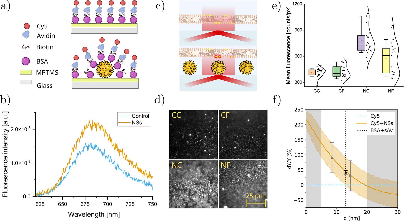
Before moving into cells, the researchers tested the design with the fluorescent molecule Cyanine-5. Coupling Cy5 to nanostars increased fluorescence to 1.49 times the control level. In artificial lipid membranes, directly coupled nanostars increased fluorescence about 1.9 times.
Distance quickly weakened the effect. Adding fibronectin increased the estimated separation between Cy5 and the nanostars from about 8.5 to 14.5 nanometers, while fluorescence enhancement dropped to about 1.3 times the control.
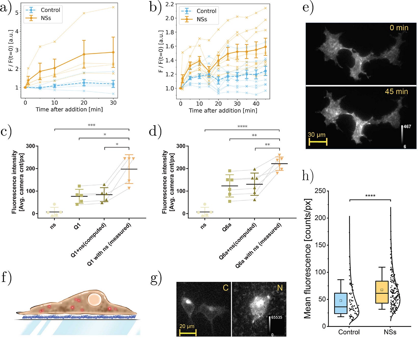
The researchers then used HEK293T cells expressing QuasAr1 or QuasAr6a. Both proteins sit in the cell membrane and report voltage through changes in fluorescence.
As nanostars accumulated around the cells, QuasAr1 fluorescence rose to 2.4 times its original level. QuasAr6a increased to 1.3 times its original fluorescence. Control tests showed that light emitted by the nanostars themselves could not explain the increase.
Placement again mattered. When researchers immobilized nanostars underneath cells and covered them with fibronectin, QuasAr6a fluorescence reached 1.69 times that of control cells. Nanostars deposited above cells produced a smaller enhancement of about 1.28 times.
The team linked that difference to spacing around the cell membrane. Their estimates placed QuasAr6a about 13.5 nanometers from nanostars beneath cells and about 16 nanometers from particles deposited above them.
Brightness was only half the surprise. Voltage-clamp experiments showed that the nanostars also changed how rapidly QuasAr6a reacted to electrical shifts.
Researchers moved membrane voltage from minus 70 to plus 30 millivolts while recording fluorescence. The protein’s upward response rate increased from about 230 per second to 1,300 per second. Its downward response accelerated from roughly 270 to 930 per second.
That improvement came with a trade-off. Relative voltage sensitivity fell from 0.37 to 0.11, a 71% reduction. The researchers attributed most of that decline to the higher baseline fluorescence rather than a major loss in the absolute voltage-dependent signal.
Control particles strengthened the case that plasmonic coupling caused the changes. Neither 50-nanometer gold nanostars nor 90-nanometer gold nanospheres produced the same effects. Only nanostars with the appropriate optical resonance changed QuasAr6a’s voltage response and speed.
The team also detected no significant additional heating under the experimental illumination conditions.
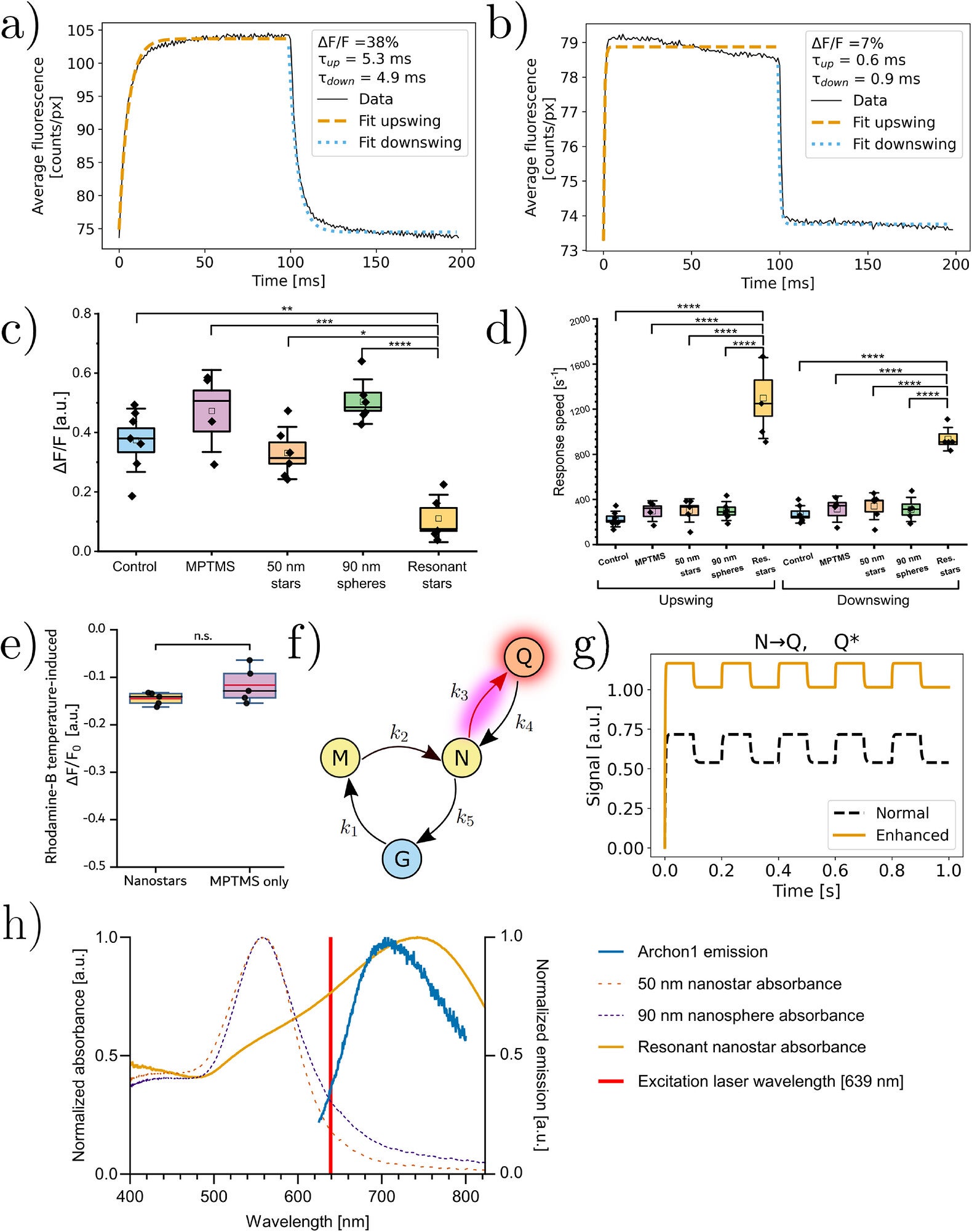
To understand the faster response, researchers built a four-state mathematical model of the QuasAr6a photocycle, the sequence of light-driven molecular states the protein moves through.
The best-fitting model required two effects at once. Plasmonic coupling increased fluorescence emission and accelerated a light-driven transition within the photocycle.
Without the nanostars, the modeled light-driven transition occurred at 443 per second. With plasmonic coupling, it rose to 1,184 per second, alongside a modeled field enhancement of 1.34.
Simply making the protein more fluorescent could not reproduce the observed acceleration. Increasing only the transition rate also produced a poorer match. The combined result suggests that the nanostars influenced both absorption into the fluorescent state and emission from it.
Their optical resonance supported that interpretation. The effective nanostars had a broad resonance peaking near 740 nanometers, overlapping both the 639-nanometer excitation light and the protein’s roughly 660-to-800-nanometer emission range.
The experiments demonstrate that plasmonic enhancement can work in a complicated mammalian cell environment without positioning every nanoparticle with molecular precision.
But the researchers could not determine the exact location of every nanostar. Some may have entered cells, and particles exposed to culture medium may have acquired a coating of proteins. Individual QuasAr6a molecules also likely experienced different coupling strengths.
The work therefore does not yet establish how the method would perform in neurons inside living brain tissue. The reported electrophysiological response rates were averaged across cells, rather than measured as identical nanoscale interactions for every protein.
Those limitations matter because brightness, response speed and voltage sensitivity all influence whether a fluorescent sensor can resolve rapid electrical events.
Genetically encoded voltage indicators have traditionally been improved by changing the proteins themselves. The nanostar experiments add another engineering route: changing the physical optical environment around an already engineered protein.
For QuasAr6a, that approach increased both fluorescence and response speed, two properties that can be difficult to improve together through genetic engineering alone.
Faster voltage reporting could help researchers measure brief electrical events more precisely if the approach can eventually be adapted to relevant neural preparations. At the same time, the loss in relative voltage sensitivity shows that making a sensor brighter does not automatically make every aspect of its performance better.
The broader result is that a nearby nanostructure can modify a functional protein’s photophysics while the protein remains embedded in a living mammalian cell. Future work can test whether tuning nanoparticle resonance, spacing and placement can better balance brightness, speed and sensitivity for biological measurements.
These resources explore the development of fast voltage indicators and the optical principles that allow nanostructures to enhance fluorescent biological measurements.
Video-based pooled screening yields improved far-red genetically encoded voltage indicators: This work introduced the screening method used to develop QuasAr6a and QuasAr6b, producing improved far-red voltage indicators for cultured neurons and live mouse brains. (Nature Methods, 2023)
Probing neuronal activity with genetically encoded calcium and voltage fluorescent indicators: This review examines recent advances in fluorescent calcium and voltage indicators and explains their role in recording neural activity at cellular and subcellular scales. (Neuroscience Research, 2025)
Seeing the Spikes: The Future of Targetable Synthetic Voltage Sensors: This perspective compares modern voltage-sensitive dyes with genetically encoded voltage indicators and discusses the remaining challenges for fast optical measurements of neuronal electrical activity. (ACS Chemical Neuroscience, 2025)
Plasmonic nanoparticle sensors: current progress, challenges, and future prospects: This broad review explains how nanoparticle shape, resonance and enhanced electromagnetic fields can improve fluorescence and other optical sensing methods. (Nanoscale Horizons, 2024)
Nanoplasmonic optical antennas for life sciences and medicine: This review describes how metallic nano-antennas can interact with biological systems for molecular imaging, cellular investigation and other life-science applications. (Nature Reviews Materials, 2018)
Research findings are available online in the journal Advanced Materials.
The original story “Gold nanostars make voltage-sensing proteins brighter and several times faster” is published in The Brighter Side of News.
Like these kind of feel good stories? Get The Brighter Side of News’ newsletter.
The post Gold nanostars make voltage-sensing proteins brighter and several times faster appeared first on The Brighter Side of News.


Leave a comment
You must be logged in to post a comment.