A jellyfish no wider than a dime can do something human skin cannot. When its surface is torn, the wound can close in minutes, often without leaving any scar at all. That quick repair has made Clytia hemisphaerica, a transparent marine animal, an unusually clear window into one of biology’s oldest problems: how living tissue decides exactly how to heal.
Jocelyn Malamy, an associate professor of Molecular Genetics and Cell Biology at the University of Chicago, first saw that process in real time about a decade ago at the Marine Biological Laboratory. Working with medusae supplied by Evelyn Houliston’s lab at the Marine Observatoire in Villefranche, she watched cells at the edge of a wound seem to walk toward one another.
The sight was striking partly because Clytia heals so fast. Small wounds can shut within minutes, and larger injuries in less than an hour. Unlike mammalian wounds, the process does not appear to produce scar tissue. “Healing in the jellyfish looks more like embryonic healing, which is scar-free,” Malamy said.
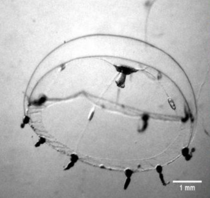
That speed alone would make the jellyfish useful. Its other advantage is simplicity. The medusae are transparent, and their outer surface is made of a single epithelial cell layer that can be watched in live animals at high resolution. They also lack several features that complicate wound studies in mammals, including vasculature, inflammation pathways, and migratory fibroblasts.
That leaves researchers with a cleaner view of epithelial repair itself. Epithelial cells cover the body’s surfaces, from skin to the lining of the gut, so they are a central focus in wound-healing research. Malamy’s latest paper, published in Molecular Biology of the Cell, uses Clytia to tackle a long-running problem in the field: why different wounds, in different systems, seem to close by different mechanisms.
In many studies, small wounds have been attributed to crawling cell extensions called lamellipodia, or to a tightening actin “purse string,” or to some combination of both. Large wounds are often linked to collective cell migration. What has been missing is a single organism where many wound types can be compared under the same conditions.
Malamy’s work argues that Clytia follows one coherent decision tree across wound sizes. The key factor is whether cells have access to exposed basement membrane, the protein sheet beneath the epithelium.
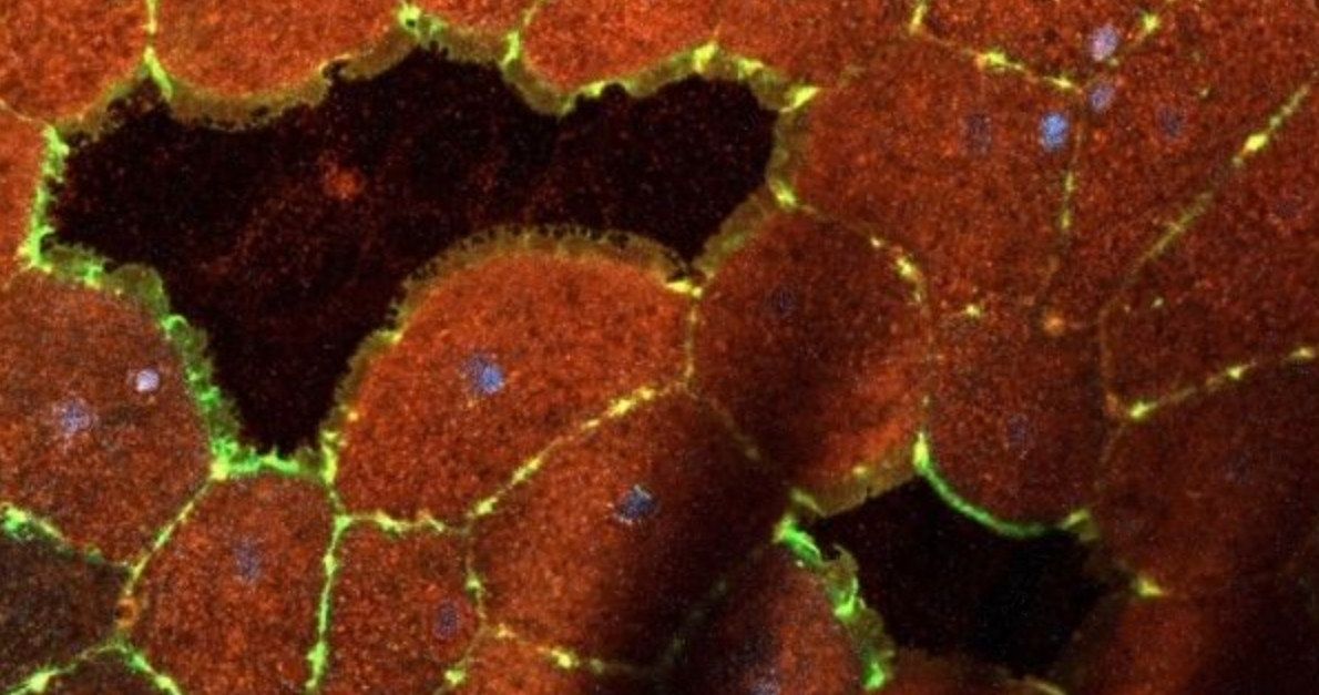
The first response is the formation of lamellipodia, which Malamy called “foot-like feelers that are actin-rich extensions of the cell.” These structures reach into the gap and crawl over the basement membrane, dragging the cell body behind them.
Her study shows that lamellipodia appear not only in larger wounds at the edge of the tissue, but even in tiny micro-wounds that cut through or between single cells. In wounds inside a single cell, the protrusions can arise from membranes internal to that cell, a result the paper describes in detail for the first time.
Those protrusions behave with surprising precision. When lamellipodia from the same cell meet, they fuse. When they come from neighboring cells, they stay separate and rebuild the boundary between cells. That suggests the tissue can distinguish “self” from “non-self” even during extremely rapid repair.
As lamellipodia move forward, a second structure forms behind them: an actomyosin cable. At first, the cable does not contract. It waits.
Then the system changes state. Once lamellipodia have covered the available basement membrane, or can no longer move because the basement membrane is damaged or blocked by debris, the actomyosin cable tightens. That contraction pulls cells together and helps expel damaged material from the wound.
The same pattern appears again and again. In micro-wounds, lamellipodia and actomyosin contraction can each close a wound on their own, making the two mechanisms partly redundant. In somewhat larger wounds, both are needed. Lamellipodia stretch cells across the gap, then the cable contracts to finish the job.
If the basement membrane is damaged, the cable becomes even more important. “If the lamellipodia have run into some debris or a tear in the basement membrane, and they can’t go any further,” Malamy said, “the actin cable can pull the cells over the basement membrane damage and also expel wound debris.”
Large wounds add another step. If the gap is too wide for lamellipodia to bridge, the epithelial sheet begins moving as a group. “The entire sheet of epithelium lifts itself up and starts walking,” Malamy said. Once the leading cells finally meet across the gap, healing returns to the same basic sequence seen in smaller injuries.
The team also found evidence that this group movement is likely triggered by mechanical signaling rather than simply by chemicals spilling from damaged cells. In large wounds, actin relocalization and protrusive behavior spread from the wound edge backward through multiple rows of cells. But when dead cells covered the basement membrane and blocked stable forward movement, that wave was greatly reduced.
The basement membrane turns out to be more than a passive surface. In this model, it acts like a decision point. As long as exposed basement membrane is available, lamellipodia remain stable and keep moving. When that surface is gone, either because the gap has been covered or because the path is blocked, lamellipodia retract and contraction begins.
That helps explain why the wound-healing literature has often seemed contradictory. Different wound types, different experimental methods, and different kinds of tissue may expose or damage the basement membrane in different ways. What looks like a contest between crawling and contraction may instead be a sequence controlled by the condition of the tissue underneath.
Malamy called it “a truly elegant mechanism where the system can rapidly adapt to heal all the kinds of wounds that might occur in nature.”
Her next step is to look beyond closure itself. Pulling cells over a damaged basement membrane may restore the surface, but it does not solve everything. “It’s great that you can heal a wound by dragging the cells over it,” she said, “but at some point, a damaged basement membrane has to get fixed.”
That remains unclear not just in jellyfish, but across wound-healing systems more broadly.
The findings do not point to an immediate medical treatment, but they sharpen a basic question at the heart of wound biology: what tells tissue whether to crawl, contract, or move collectively?
By isolating that decision in a simple, transparent animal, the work offers a clearer framework for studying epithelial repair in more complex organisms. It also suggests that some disagreements in the field may reflect differences in basement membrane damage rather than wholly different healing programs.
If that idea holds up in vertebrate tissues, it could help researchers rethink how wounds are modeled in the lab and how scar-free repair might one day be encouraged.
Research findings are available online in the journal Molecular Biology of the Cell.
The original story “Giant dark matter ‘sheet’ may shape galactic motion in the Milky Way” is published in The Brighter Side of News.
Like these kind of feel good stories? Get The Brighter Side of News’ newsletter.
The post Jellyfish can heal wounds in minutes. Could humans be next? appeared first on The Brighter Side of News.


Leave a comment
You must be logged in to post a comment.