Obesity does not just enlarge fat stores, it appears to rework the body’s wiring and immune landscape in ways scientists have struggled to see. Now, whole-body mouse maps point to damaged facial sensory nerves, and to inflammatory hotspots with wider implications.
Obesity is easy to spot on a scale. What has been much harder to see is how deeply it reshapes the body beneath the surface. This ranges from immune-cell buildups in swollen tissue to subtle damage in nerves that help animals sense the world around them.
That blind spot may now be narrowing.
A research team led by Prof. Ali Ertürk of Helmholtz Munich and Ludwig Maximilians University Munich (LMU) has developed a whole-body imaging and analysis system called MouseMapper. This deep-learning framework can scan intact transparent mice and automatically map nerves, immune cells, organs, and tissues across the body. In obese mice, the approach turned up widespread inflammatory changes. In addition, it revealed an unexpected form of nerve remodeling in the face.
The work pushes past a long-standing problem in obesity research. Scientists have had ways to examine selected organs in detail, but not to track disease-linked changes across an entire intact body at high resolution. That matters in obesity, which raises the risk of type 2 diabetes, cardiovascular disease, stroke, peripheral neuropathies, and many cancers. Obesity also affects far more than fat metabolism alone.
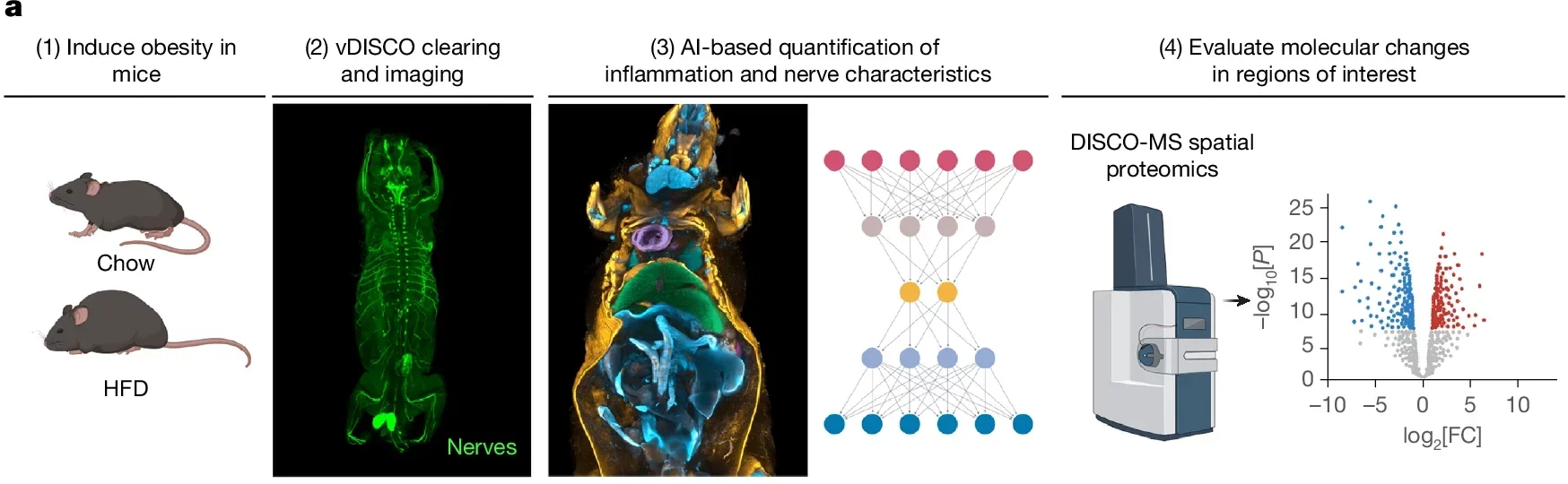
To build these maps, the researchers used mice engineered so that peripheral nerves or monocytes and macrophages glowed under fluorescent light. They then applied tissue-clearing methods that made the animals transparent while preserving those signals.
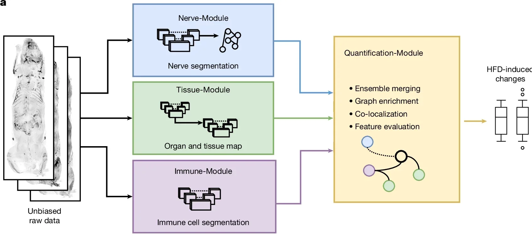
With light-sheet fluorescence microscopy, the team captured three-dimensional images of whole mouse bodies. These images included datasets containing tens of millions of cellular structures. MouseMapper then analyzed those scans with three linked modules: one for nerves, one for immune cells, and one for mapping those findings onto 31 organs and tissue types.
“MouseMapper is built on a foundation model, which means it generalizes far beyond the data it was originally trained on,” said Ying Chen, co-first author of the study.
That generalizability is one of the main technical claims here. The nerve-analysis module was built from VesselFM, a pre-trained foundation model originally designed for blood vessel segmentation. Because blood vessels and nerves share elongated branching structures, the team reasoned that those learned features could transfer. After fine-tuning, the model outperformed systems trained from scratch, reaching a voxel Dice score of 0.7494 for nerve segmentation. The immune module also beat earlier methods, with a voxel Dice score of 0.7878.
The framework did more than produce attractive maps. It let the team compare animals without choosing target regions in advance. This means disease hotspots could emerge on their own.
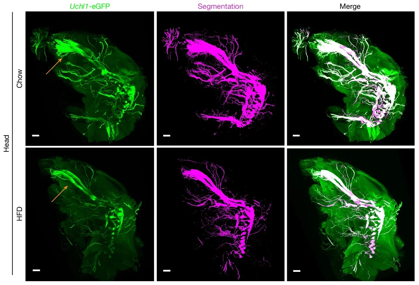
One of the clearest patterns appeared in the head.
Mice fed a high-fat diet for 16 to 18 weeks developed obesity, gained adipose tissue, and showed impaired insulin response. Across the whole body, their nerve density fell even when total nerve voxels stayed similar. This suggests that as body size expanded, innervation did not keep pace. Adipose tissue followed the same pattern: more total nerve voxels because the tissue was bigger, but lower nerve density overall.
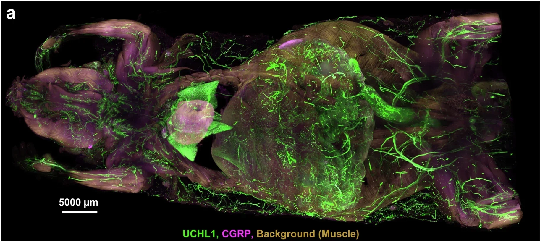
The most striking changes showed up in the infraorbital nerve, part of the trigeminal system that helps relay facial sensory information and supports whisker-based exploration. In obese mice, that nerve had sharply reduced complexity. The number of nerve endings dropped by 60.7 percent, edges by 57.8 percent, and vertices by 57.6 percent. Nerve thickness, however, did not significantly change. This points less to wholesale collapse than to defects in axonal extensions branching away from the ganglia.
The animals also behaved differently. In whisker-stimulation tests, obese mice showed a weaker response, linking the structural damage to sensory impairment.
That combination, anatomy plus function, gave the finding extra weight.
“We revealed previously unknown structural and molecular changes in the trigeminal ganglion and its facial branches, and the same molecular signature was conserved in human tissue. This kind of finding simply cannot emerge from studying one organ at a time,” said Dr. Doris Kaltenecker, senior scientist at the Institute for Diabetes and Cancer at Helmholtz Munich and first author of the study.
The team then looked upstream, at the trigeminal ganglion, where the cell bodies of those sensory neurons sit.
Using spatial proteomics, they analyzed trigeminal ganglia from chow-fed and high-fat-diet mice and identified more than 6,000 proteins in each sample. Of those, 230 were differentially regulated, including 67 upregulated and 163 downregulated proteins. Pathway analysis pointed to changes involving actin cytoskeleton regulation, RHO GTPase effectors, axon guidance, complement and coagulation cascades, ERBB signaling, and sphingolipid signaling.
Several SERPIN-A family proteins were downregulated, including molecules tied to limiting inflammation-related tissue damage. The team also validated selected signals with western blotting, including reduced SERPINA1 expression, ERK activation, and increased SEPTIN7 expression.
Then came the translational test. The researchers examined post-mortem trigeminal ganglia from lean people with body mass index below 25. They also studied people with obesity whose BMI exceeded 30. Proteomic profiling showed obesity-linked changes in pathways related to axon guidance, neurodegeneration, and actin cytoskeleton regulation, mirroring the mouse findings.
That does not prove the same symptoms occur in people, but it does suggest the molecular signature is not confined to a mouse model.
MouseMapper also gave the team a full-body view of immune activity.
In obese mice, Cd68-positive immune cells appeared throughout the body, especially in the liver and visceral adipose tissue. The researchers sorted these immune clusters by size, from small groups of up to six cells to large aggregates of more than 60 cells. Larger clusters can reflect a more activated and pro-inflammatory state.
The pattern shifted with obesity. Small-cluster proportions fell in the liver, visceral fat, and stomach. Medium clusters rose in the liver and visceral fat. Large clusters increased in several areas, including subcutaneous fat, visceral fat, muscle, stomach, abdominal wall, adrenal glands, Peyer’s patches, and vesicular gland.
Higher-resolution and multiplex labeling added more detail. In visceral fat, macrophage-rich clusters often sat near T cells, natural killer cells, and endothelial cells. This suggests perivascular immune hubs rather than isolated cell piles.
The body-wide pattern was not random. It was regional, uneven, and tied to tissue context.
The system is powerful, but not all-seeing.
The standard whole-body imaging setup could not fully resolve the thinnest axons and subcellular features. A higher-resolution 4x method improved detection and cut acquisition time from nearly two weeks to about 20 hours. However, it also drove data loads up to 50 terabytes per mouse. The authors also note that full generalizability across different imaging modalities is still out of reach, so new datasets may require further fine-tuning.
Even so, the broader message stands. Obesity did not merely enlarge fat depots in these animals. It altered nerve organization, immune-cell clustering, lymph node mass, liver volume, and molecular pathways. This played out across connected systems rather than inside one isolated organ.
MouseMapper gives researchers a way to find disease hotspots across an intact body instead of guessing where to look first. In obesity, that approach uncovered facial sensory nerve remodeling and widespread inflammatory changes. These changes may have been missed in organ-by-organ studies.
The platform could also be adapted to other conditions that spread across systems, including diabetes, cancer, neurodegeneration, and autoimmune disease. By pairing whole-body maps with molecular analysis, scientists may be able to identify earlier structural warning signs.
They can also narrow down therapeutic targets and reduce the number of physical experiments needed to test new ideas.
Research findings are available online in the journal Nature.
The original story “Obesity damages your face as well as nerves across the whole body, AI finds” is published in The Brighter Side of News.
Like these kind of feel good stories? Get The Brighter Side of News’ newsletter.
The post Obesity damages your face as well as nerves across the whole body, AI finds appeared first on The Brighter Side of News.


Leave a comment
You must be logged in to post a comment.