Pale, fingerlike structures rising from tree roots in southwestern China have revealed an unexpected giant among fungi. Named Dianjunus rex, the newly described species produces fleshy reproductive structures reaching 50 centimeters in total height. The largest reported specimen weighed 2.2 kilograms, roughly 4.9 pounds.
Its size was only the beginning of the surprise. Genetic comparisons and microscopic examinations placed the fungus on a distinct evolutionary branch, supporting a new genus and family. The findings appeared in Frontiers in Fungal Biology.
The team included scientists from the Kunming Institute of Botany, Chinese Academy of Sciences, and collaborating institutions. Corresponding author Indunil C. Senanayake works at the institute’s Germplasm Bank of Wild Species. He described the discovery as a rare opportunity to investigate the evolution of extreme fungal forms.
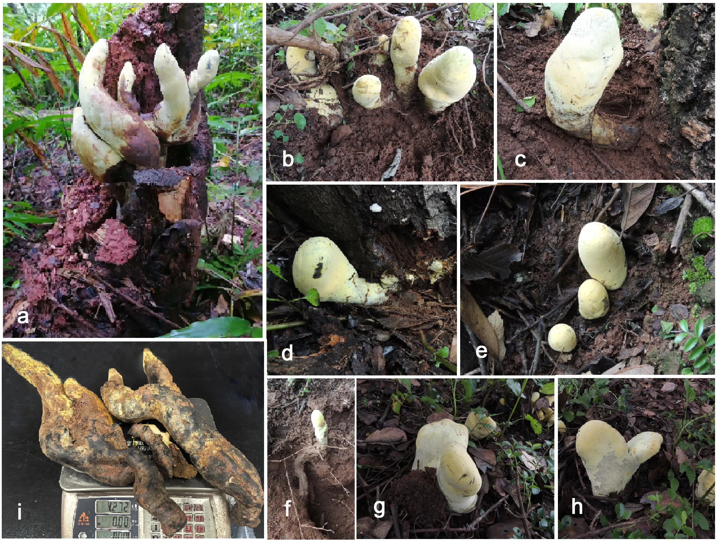
Collections came from several locations in Yunnan Province. Fieldwork near Damaidi took place in August and September 2024, followed by collections near Nanbang and Yipinglang in July 2025. The sites included coniferous forests and tropical rainforest, showing that the fungus occurs across contrasting environments.
Some specimens were associated with Chinese cork oak, Quercus variabilis. Others came from another oak or an unidentified decaying tree. The evidence therefore does not restrict the species to a single tree host.
The structures are called stromata, tissues containing the fungus’s spore-producing chambers. Their forms range from thick cylinders to flattened, palm-shaped growths, sometimes branching near the base or top. Young specimens appear whitish, becoming yellow to darker yellow as they mature.
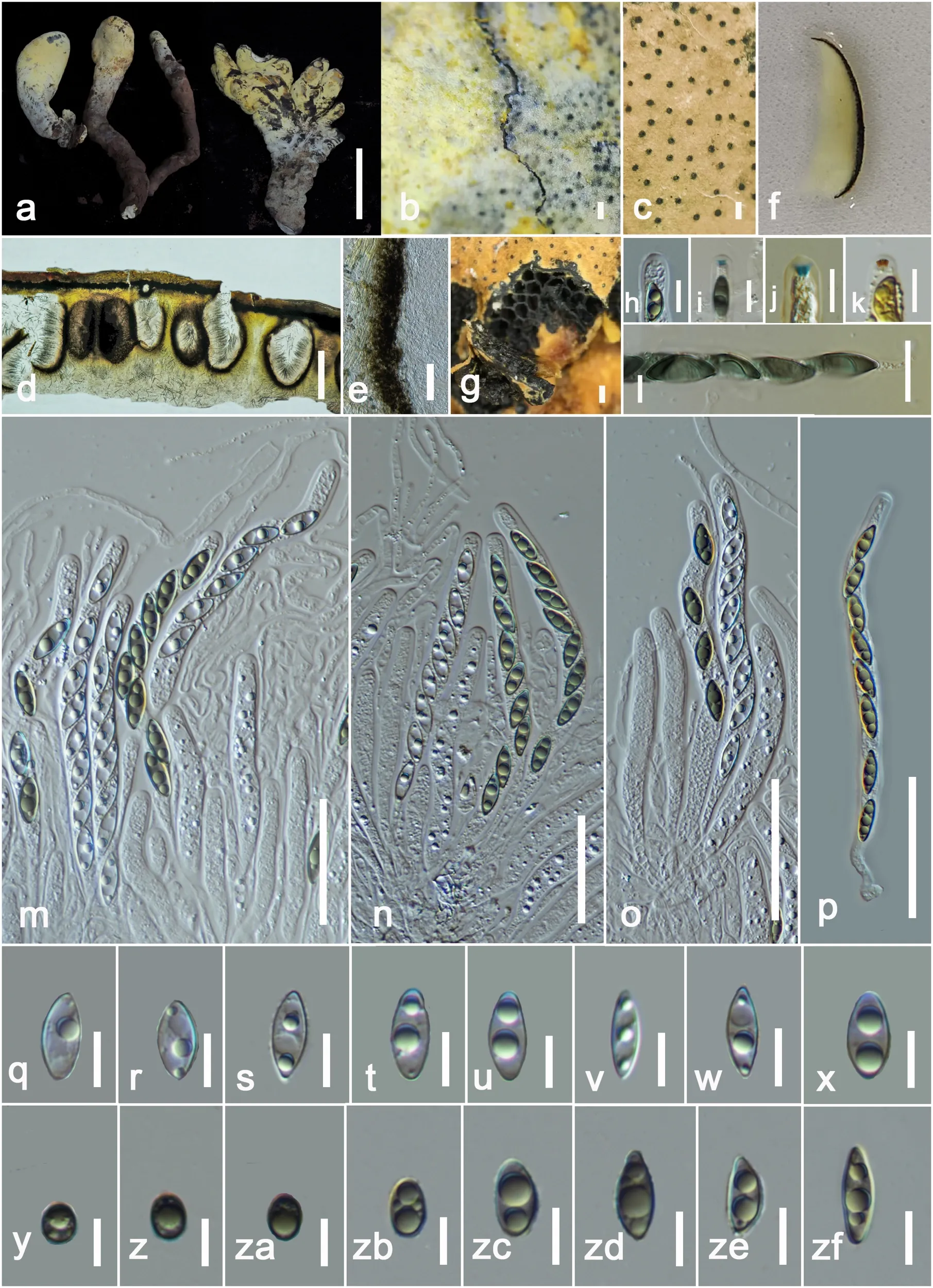
Ascomycota takes its name from the microscopic sacs, called asci, in which sexual spores develop. In this species, those sacs sit inside chambers embedded in the larger tissue. Tiny openings appear as black dots on its surface, revealing the reproductive machinery beneath the pale coating.
The height measurement needs context. The formal description gives a total height of 20–50 centimeters, including a substantial buried portion, rather than a half-meter column entirely above ground. Its aerial fertile region measures 10–20 centimeters high, while dark, rootlike stalks extend into the soil.
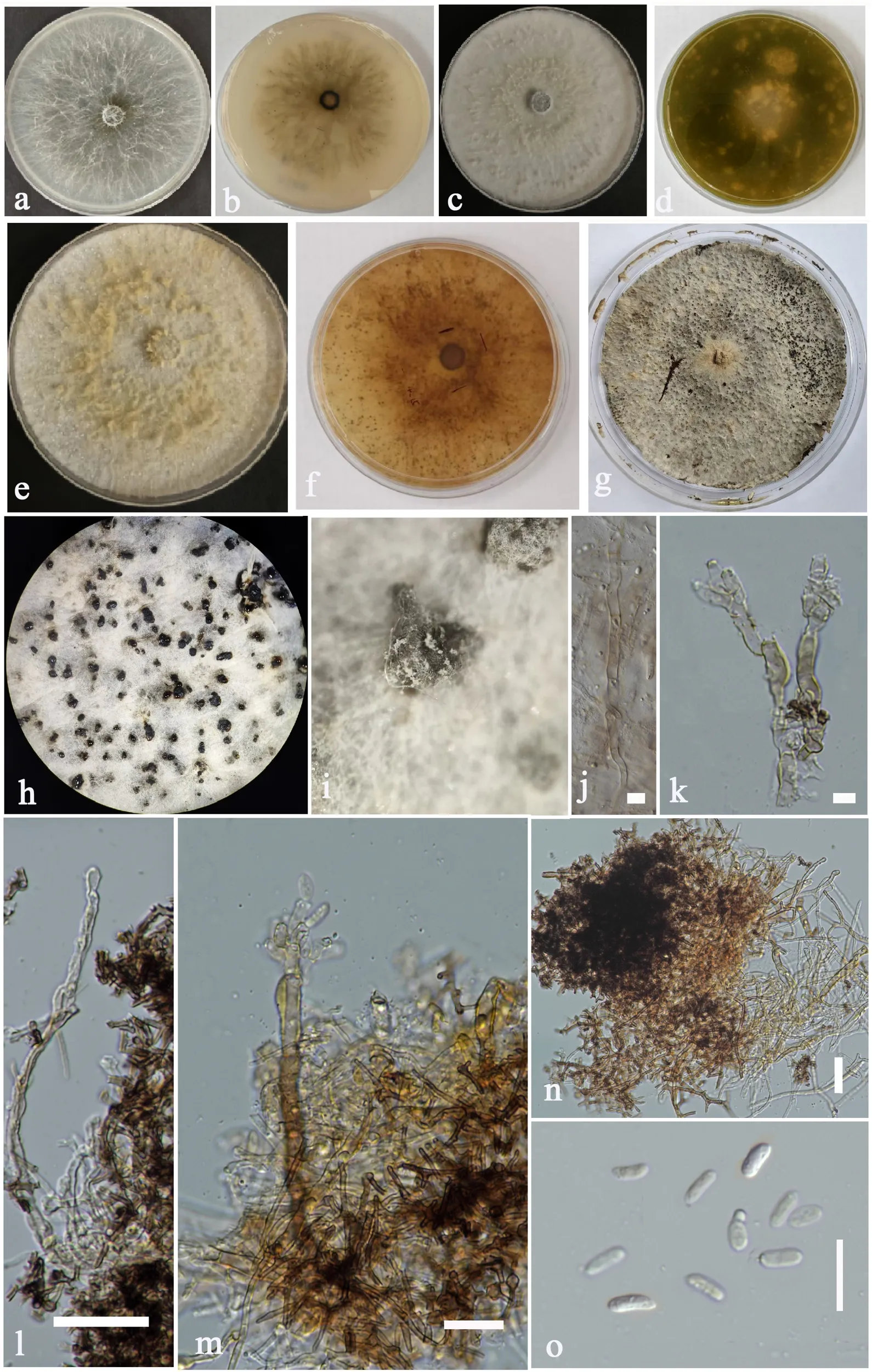
The researchers preserved specimens in the Kunming Institute of Botany’s fungarium, creating reference material for future comparisons. They also cultured individual spores and examined the resulting growth. Those cultures allowed them to study features that would be difficult to distinguish in a forest specimen.
Under the microscope, the fungus revealed several useful identifying traits. Its brown spores have a conspicuous S-shaped slit, and its spore-containing sacs possess a distinctive apical structure. A persistent powdery, fibrous coating also distinguishes the larger reproductive tissues.
An older described fungus, Squamotubera le-ratii, initially offered a possible comparison. However, its surface, internal tissues and other recorded characteristics differed from the Yunnan specimens. That comparison helped show why a superficial resemblance was insufficient to assign the collections to an existing genus.
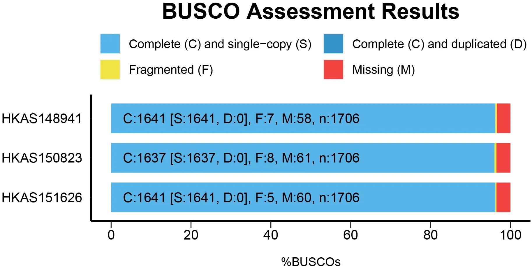
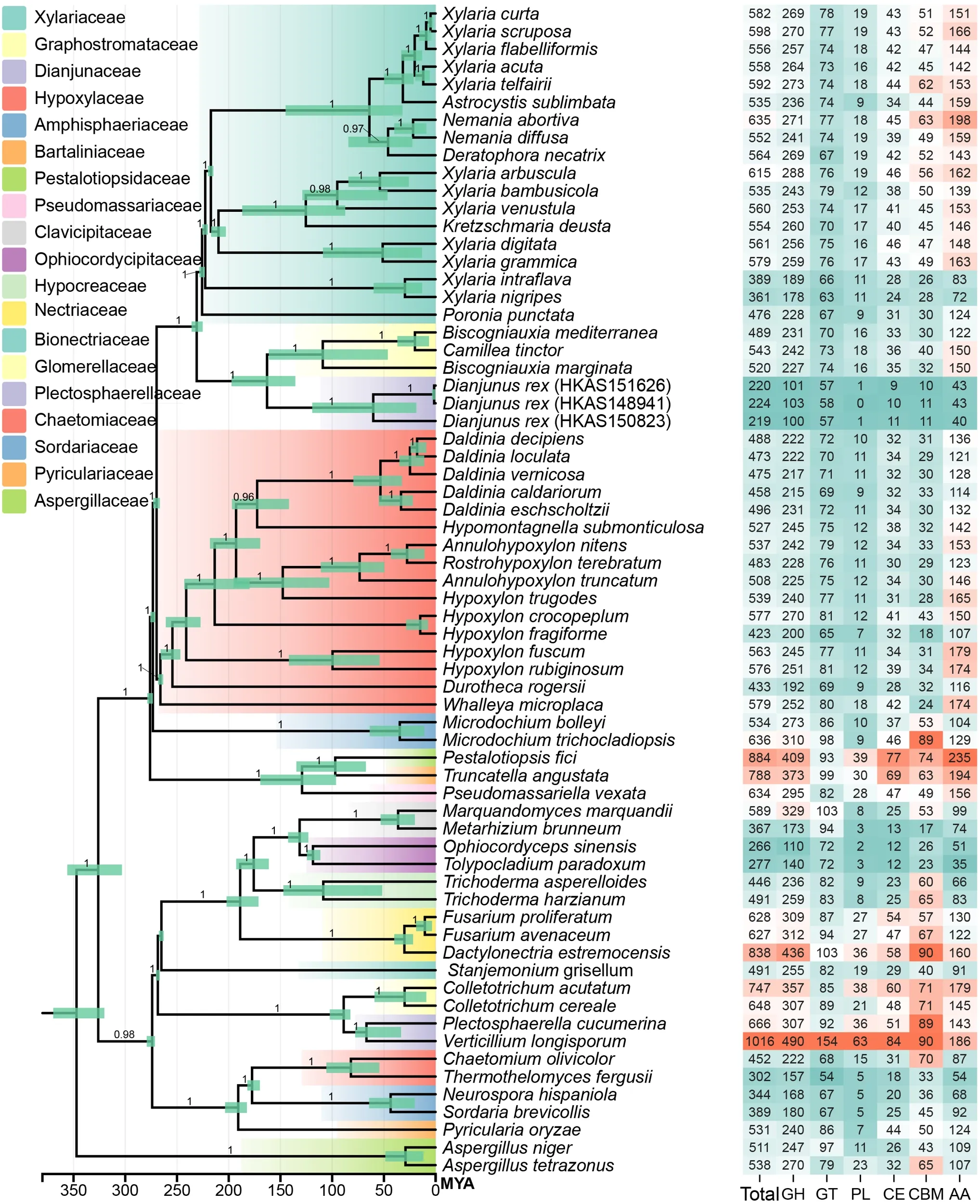
The team compared four genetic regions across a dataset containing 202 strains. A separate genome-scale analysis combined three newly sequenced samples with data from 63 other fungal species. Multiple analytical approaches consistently placed the new lineage beside the family Graphostromataceae.
Together, the genetic and anatomical evidence supported naming the species Dianjunus rex and establishing the family Dianjunaceae. The genus name references Yunnan’s ancient Dian kingdom and the Chinese word for fungus. The Latin rex, meaning king, acknowledges its imposing form.
The paper describes the species as producing the largest documented stromata within Ascomycota. That broad claim deserves qualification, however, because the same paper cites a heavier example from another species. A Kenyan specimen of Engleromyces goetzei reportedly weighed approximately 3.2 kilograms and measured nearly 30 centimeters across.
The strongest comparison therefore concerns the new fungus’s exceptionally large upright structures, rather than an unqualified weight record. Height, mass and volume describe different aspects of size. The study’s 2.2-kilogram maximum also refers to a large, two-lobed specimen, while the formal description lists smaller weights for other stromata.
Nor does the measurement describe the size of an entire fungal organism. The visible reproductive structure represents only one part of a fungus’s growth. The study did not map the full extent of its underground network or establish a record across all fungi.
Senanayake suggested that greater height might help spores escape surrounding vegetation and forest debris. That potential advantage would have to compensate for the energy needed to build large reproductive tissues. The research did not directly measure spore dispersal or test whether taller specimens reproduce more successfully.
The researchers also used genetic differences and calibrated evolutionary models to estimate when Dianjunaceae separated from related lineages. Their central estimate was approximately 65 million years ago. That falls close to the mass extinction about 66 million years ago that eliminated non-avian dinosaurs and transformed ecosystems.
Such disruption could have created opportunities for fungi associated with dead plant material. Forest destruction would have changed the availability of potential food and habitats. Senanayake proposed that these conditions might have favored some wood-associated fungal lineages.
The estimate nevertheless spans approximately 50–80 million years, covering times both before and after the extinction. It cannot establish that the family originated precisely one million years afterward. It also dates an evolutionary split, rather than the age of a living specimen or the first appearance of its giant form.
Genome comparisons uncovered an unexpected feature beneath the fungus’s large exterior. The three samples contained approximately 219–224 genes for carbohydrate-active enzymes, substantially fewer than several related decomposer fungi. Genes encoding one group of plant-wall-degrading enzymes, pectin lyases, were almost entirely absent.
Those patterns led the authors to propose a possible change in lifestyle. The fungus might first inhabit living plant tissues without obvious symptoms, then shift toward decomposition after its host dies. Its observed fruiting after host mortality is consistent with that idea, but does not establish the complete sequence.
Finding reproductive structures beside dying or dead trees also leaves an important question unresolved. That association alone does not show whether the fungus contributed to the trees’ decline. Establishing its relationship with living hosts would require further investigation.
The team further suggested that reduced investment in some degradative enzymes could accompany greater allocation to building fungal tissue. Testing that explanation will require measurements beyond gene inventories. For now, the discovery provides preserved specimens, cultures and genome data with which to investigate how this unusual fungus lives and grows.
These resources explore fungal classification, flexible lifestyles and the ecological changes surrounding ancient mass extinctions.
Exploring the Xylariaceae and its relatives: Reviews the diversity and biological features of the broader fungal group containing large stromata-producing species. (Botanical Studies, 2023)
Large-scale genome sequencing of mycorrhizal fungi provides insights into the early evolution of symbiotic traits: Shows how genome comparisons reveal changes in fungal lifestyles and enzyme repertoires. (Nature Communications, 2020)
Time to re-think fungal ecology? Fungal ecological niches are often prejudged: Discusses evidence that fungi can occupy multiple ecological roles rather than fitting one fixed category. (New Phytologist, 2018)
Fungal proliferation before and after the Cretaceous–Paleogene mass extinction event in North America: Examines fossil evidence for fungal abundance around the extinction boundary. (PNAS, 2026)
Fungal Diversity Revisited: 2.2 to 3.8 Million Species: Explains the methods and uncertainties involved in estimating global fungal diversity. (Microbiology Spectrum, 2017)
Research findings are available online in the journal Frontiers in Fungal Biology.
The original story “Scientists discover ‘D-Rex,’ a giant fungus with a 65-million-year evolutionary history” is published in The Brighter Side of News.
Like these kind of feel good stories? Get The Brighter Side of News’ newsletter.
The post Scientists discover ‘D-Rex,’ a giant fungus with a 65-million-year evolutionary history appeared first on The Brighter Side of News.


Leave a comment
You must be logged in to post a comment.