Tiny packages naturally released by human cells reached the brain after being delivered through the nose and protected memory in mice with Alzheimer’s-like disease. The same vesicles also supported neurons grown from cells donated by people with Alzheimer’s.
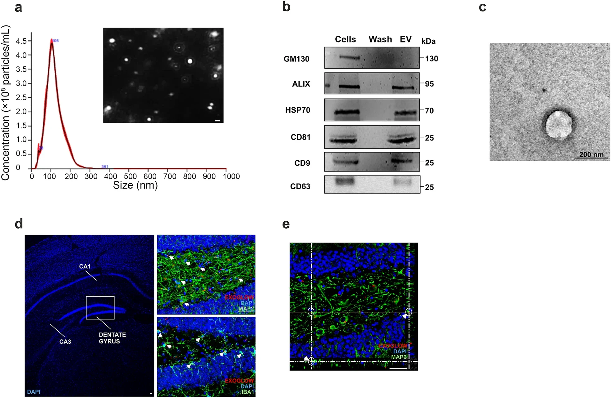
The experimental treatment uses extracellular vesicles, or EVs, produced by human amniotic membrane mesenchymal stromal cells. These membrane-bound particles carry proteins, regulatory RNAs and other biological material between cells.
Researchers from Università Cattolica del Sacro Cuore, Fondazione Policlinico Universitario A. Gemelli IRCCS and the E. Menni Research Center studied whether those vesicles could influence several Alzheimer’s-related processes at once.
The results link improved memory with changes in inflammation, amyloid-beta and proteins needed for communication between neurons. But the researchers emphasize that the work does not yet establish a treatment for people.
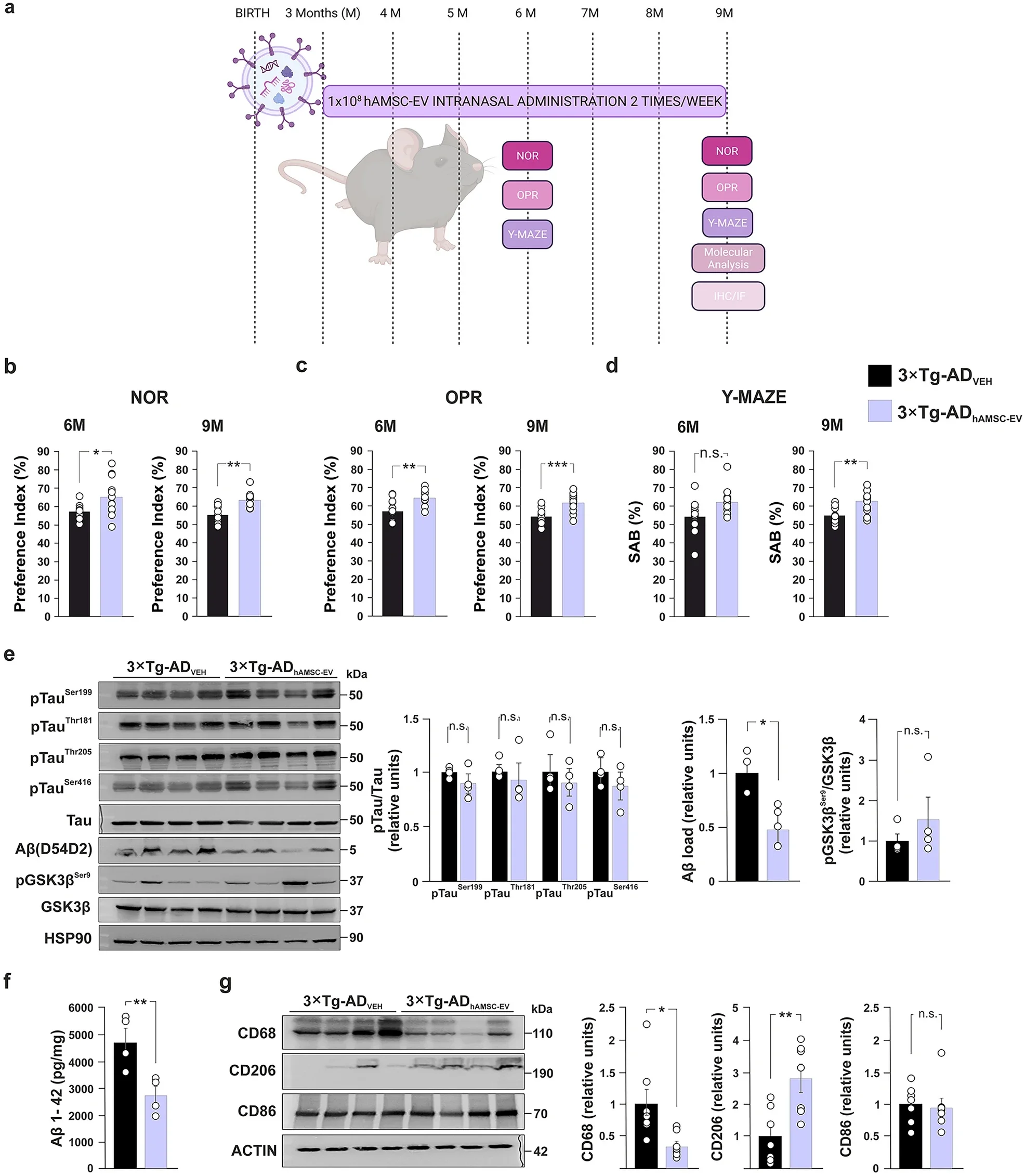
The main experiments used female 3×Tg-AD mice, which develop several Alzheimer’s-related abnormalities with age. Treatment began when the animals were 3 months old, before cognitive problems appeared, and continued until 9 months.
Twice each week, mice received about 100 million vesicles in 4 microliters of solution through the nose. Control animals received saline.
Fluorescent labeling showed that the vesicles reached the hippocampus, a brain region important for memory. They were associated with both neurons and microglia, immune cells that help monitor conditions within the brain.
Memory testing revealed differences after both three and six months of treatment. In novel object recognition, treated mice scored 65.19% after three months compared with 57.29% in controls. After six months, the scores were 63.29% and 55.23%, respectively.
Treated animals also performed better in object place recognition and a Y-maze test of spatial working memory.
The behavioral changes appeared alongside measurable differences in the brain.
Hippocampal amyloid-beta fell by 53% in treated 9-month-old mice. Levels of Aβ1-42 measured 2,743.7 picograms per milligram in treated animals compared with 4,712.5 in controls.
The treatment did not significantly change tau phosphorylation at the sites examined. Researchers also found no significant change in GSK3β phosphorylation.
That contrast is important because the treatment did not uniformly alter every major Alzheimer’s-related process.
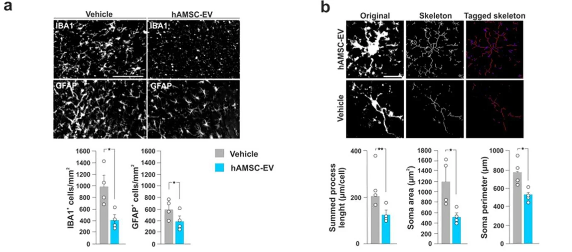
Brain inflammation changed more substantially. Treated mice had fewer Iba1-positive microglia and GFAP-positive astrocytes in vulnerable regions. Microglia also became less branched and structurally complex.
Rather than describing the treatment as simply suppressing immune activity, the researchers interpret the findings as a remodeling of the inflammatory environment surrounding neurons.
The vesicles also affected proteins involved in neuronal plasticity and communication.
Levels of BDNF, GluA1 and ARC increased after treatment. These proteins are connected with synaptic function and the ability of neuronal networks to adapt.
Laboratory experiments with activated microglia provided another clue. Pretreatment with the vesicles reduced uptake of fluorescent amyloid-beta by 51.5% and reduced reactive multinucleated cells by 55.7%.
Phosphorylated NF-kB declined by 25.7%, while the marker CD206 increased by 23.6%. Together, the results pointed toward a shift in inflammatory signaling rather than a complete shutdown of microglial activity.
The team also tested a shorter intervention beginning after cognitive problems had appeared. Mice treated from 9 to 10 months improved in novel object recognition, although they did not improve in object place recognition.
That result offers preliminary evidence that the vesicles may do more than prevent early deficits, but the shorter experiment was not equally successful across memory tests.
Researchers next examined what inside the vesicles might drive these changes.
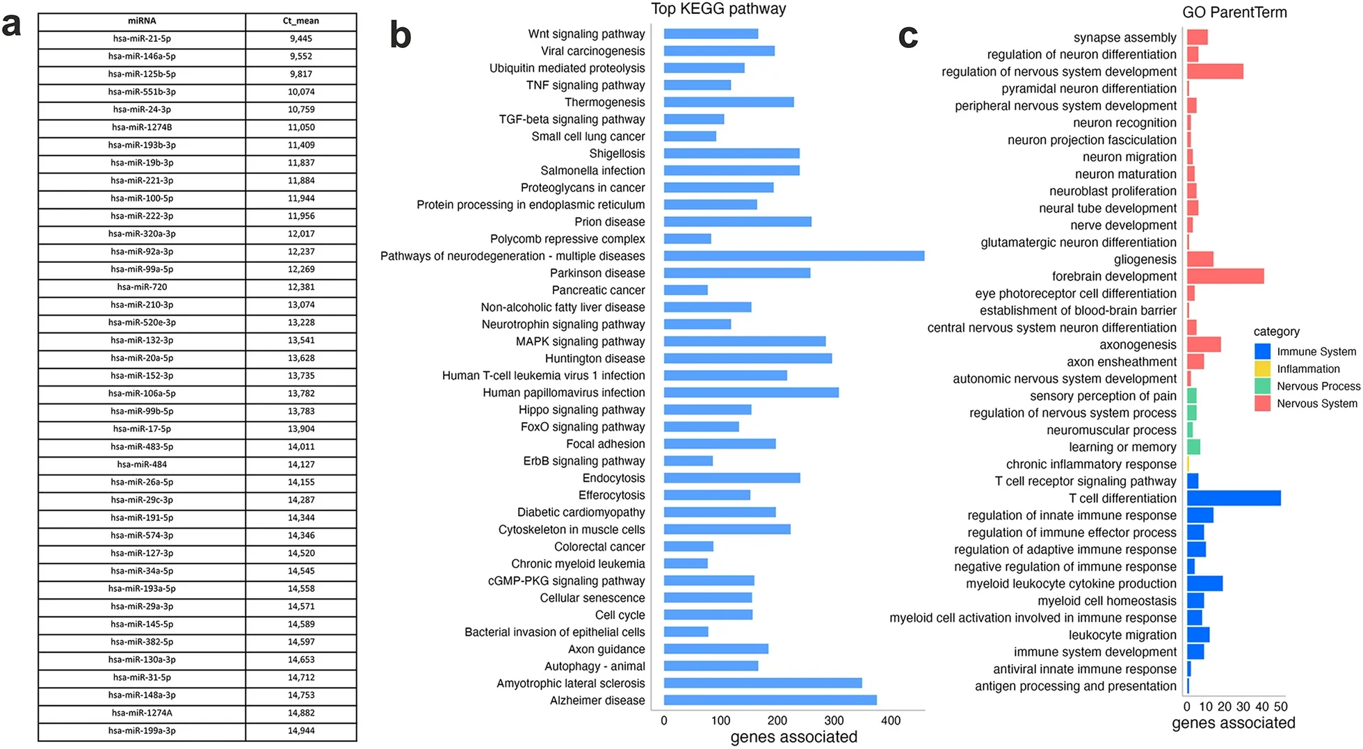
Their cargo included numerous microRNAs, small regulatory molecules that can affect gene activity. Abundant examples included miR-21-5p, miR-146a-5p, miR-125b-5p and miR-100-5p.
Bioinformatic analysis connected predicted targets of these molecules with pathways involving inflammation, nervous-system development, synapses, learning and memory.
To test whether RNA mattered directly, the researchers depleted much of the vesicles’ RNA cargo. Those altered vesicles became less effective at reducing inflammatory activation in microglia.
The experiment did not identify a single responsible molecule because many RNAs were removed together. Instead, it supports the idea that the vesicles’ regulatory RNA cargo contributes to their biological activity.
The team then turned to human cells.
Skin fibroblasts from three people with sporadic Alzheimer’s and three healthy participants were reprogrammed into induced pluripotent stem cells and then developed into glutamatergic neurons.
Untreated Alzheimer’s-derived neurons developed shorter neurites, the projections used to form connections. Vesicle treatment substantially improved neurite length and restored several synaptic proteins that were reduced in the Alzheimer’s-derived cells.
Synapsin-1 increased 131.5% relative to untreated Alzheimer’s neurons. PSD95 rose 112.5%, while synaptophysin and GluA1 also increased.
Removing RNA from the vesicles weakened several of those effects, again pointing toward the importance of their molecular cargo.
The findings combine several lines of evidence: better memory in mice, reduced amyloid, altered inflammatory activity and improved synaptic markers in both mouse brains and human-derived neurons.
They also leave major questions unanswered.
The main animal experiments involved female genetically engineered mice. Human experiments took place in laboratory-grown neurons, not in people receiving an intranasal treatment. The study also cannot yet determine which vesicle components matter most or what dose and schedule would be appropriate for humans.
“These are preclinical results that require further validation in humans and do not yet represent an available therapy for Alzheimer’s disease,” said Claudio Grassi of Università Cattolica.
The broader idea is that Alzheimer’s treatment may eventually need to address more than amyloid-beta and tau alone. This work points toward the cellular environment around neurons, including microglia, inflammatory signals and synaptic support, as another possible therapeutic target.
These studies and reviews examine how extracellular vesicles influence neuroinflammation, synapses and experimental Alzheimer’s treatment, including intranasal delivery to the brain.
Intranasal administration of neural stem cell-derived extracellular vesicles prevents cognitive decline in both male and female 3×Tg-AD mice by dampening neuroinflammation and epigenetically regulating amyloid β metabolism: Neural stem cell-derived vesicles delivered through the nose delayed cognitive decline, reduced hippocampal inflammation and decreased amyloid-beta in male and female Alzheimer’s-model mice. (Alzheimer’s Research & Therapy, 2026)
Intranasal Adipose-Derived MSC Extracellular Vesicles Confer Sustained Cognitive Improvement and Suppress Alzheimer’s Pathology in APP/PS1 Mice: Intranasal extracellular vesicles reached several brain regions and improved memory while reducing hippocampal amyloid pathology, with some benefits persisting after treatment stopped. (Biomolecules, 2026)
Extracellular Vesicles for the Treatment of Alzheimer’s Disease: A Systematic Review: This systematic review evaluates preclinical evidence for cell-derived vesicles in Alzheimer’s and highlights mechanisms, model limitations and the need for standardized EV production and testing. (Journal of Extracellular Biology, 2025)
Extracellular vesicles as therapeutic modulators of neuroinflammation in Alzheimer’s disease: a focus on signaling mechanisms: This review focuses on how extracellular vesicles may alter inflammatory signaling involving microglia, astrocytes and pathways such as NF-kB in Alzheimer’s disease. (Journal of Neuroinflammation, 2025)
Intranasal delivery of engineered extracellular vesicles loaded with miR-206-3p antagomir ameliorates Alzheimer’s disease phenotypes: Engineered vesicles carrying a microRNA-targeting molecule improved memory, neurogenesis and synaptic plasticity while reducing amyloid deposition in an Alzheimer’s mouse model. (Theranostics, 2024)
Research findings are available online in the journal Springer Nature.
The original story “Tiny extracellular vesicles delivered through the nose found to improve memory in Alzheimer’s study” is published in The Brighter Side of News.
Like these kind of feel good stories? Get The Brighter Side of News’ newsletter.
The post Tiny extracellular vesicles delivered through the nose found to improve memory in Alzheimer’s study appeared first on The Brighter Side of News.


Leave a comment
You must be logged in to post a comment.