Breathing may feel like one of the body’s most automatic routines, but no two breaths are quite the same. One inhale may arrive quickly, another slowly. An exhale can stretch out, stop briefly or carry a different amount of air than the breath before it.
The brain appears to notice those differences.
A study led by researchers at the University of California San Diego found that the detailed shape of individual breaths corresponds with the shape of neural activity recorded across parts of the human brain. Rather than responding only to breathing as a repeating rhythm, the brain appears to track changes from one breath to the next.
The findings, published in the Journal of Neuroscience, suggest that breathing and neural activity are linked on a finer timescale than traditional measures such as breaths per minute can reveal.

“Every single breath is different,” said first author Eena Kosik-Rose, a PhD student in UC San Diego’s Department of Cognitive Science. “You can pause your breathing for several seconds, take a super deep breath or have a shallow exhale. What we’re showing is that those differences in the shape of each breath are reflected in the shape of brain activity.”
Scientists have long known how brainstem networks regulate the basic mechanics of breathing. More recent work has connected respiration with higher-level functions, including attention, perception, emotion and memory.
Previous experiments have shown, for example, that performance on some tasks can change depending on whether information arrives during inhalation or exhalation. Breathing also produces rhythms in neural activity across several areas of the forebrain.
Most studies of that connection, however, have treated respiration mainly as a repeating cycle. Researchers commonly measure how strongly brain activity synchronizes with the overall frequency of breathing.
That approach can miss what happens inside individual breaths.
Human respiration is not a perfectly regular wave. Inhalation and exhalation have different durations, intensity varies, and pauses can occur between cycles. Kosik-Rose and her colleagues wanted to know whether those small differences were mirrored in neural signals.

“Our results show that the coupling between breathing and neural activity is much richer than previously appreciated,” said Bradley Voytek, a study coauthor and professor and chair of UC San Diego’s Department of Cognitive Science.
The study analyzed previously collected intracranial recordings from 16 people undergoing clinical monitoring for treatment-resistant epilepsy. Participants came from Northwestern Memorial Hospital, Children’s National Hospital and the University of Iowa Stead Family Children’s Hospital.
Electrodes implanted for clinical purposes recorded electrical activity directly from the brain. Researchers simultaneously measured respiration using nasal airflow sensors, breathing belts around the chest or abdomen, or both.
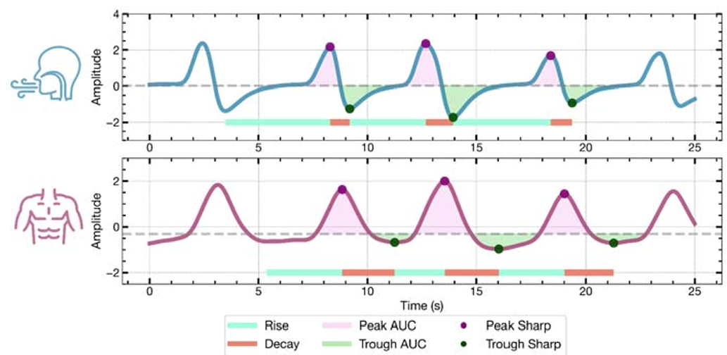
The team represented each breath as a wave and measured features including how long airflow took to rise and fall, the duration of the complete breathing cycle, the amount of airflow during inhalation and exhalation, and the sharpness of respiratory peaks and troughs.
They then paired individual respiratory cycles with neural oscillations recorded at the same time.
The analysis first identified brain channels whose activity was synchronized with breathing. Significant respiratory coherence appeared across distributed cortical and limbic regions, including areas of the frontal and temporal cortex, hippocampus, amygdala and insula.

But synchronization alone did not mean that each neural wave followed the detailed form of each breath. The researchers applied additional tests to find channels showing consistent alignment on a cycle-by-cycle basis.
After those screening steps, 18 channels across seven participants showed significant waveform coupling with nasal airflow. Only four belt-recorded channels across three participants passed the same criteria.
Within the airflow measurements, several aspects of each breath showed strong evidence of corresponding changes in neural activity. These included rise time, decay time, total cycle duration and measurements related to the amount of airflow during inhalation and exhalation.
The sharpness and symmetry of breaths did not show the same clear relationships.
The much stronger result for nasal airflow may be important. Earlier research has connected nasal breathing with neural activity through pathways involving the olfactory system, although other evidence suggests breathing can synchronize with the forebrain even when airflow bypasses the nose.
The current study cannot determine which pathway causes the effect. Breathing belts also provide a coarser measurement of chest movement than nasal sensors provide of airflow, which could partly explain the weaker belt results.
What the recordings do show is that neural activity can follow breath-to-breath variations with surprising precision.
That precision raises questions that extend beyond normal cognition. One area the team hopes to investigate involves sudden unexpected death in epilepsy, or SUDEP, in which a person with epilepsy dies suddenly.
Study coauthor Brian Dlouhy, a neurosurgeon at the University of Iowa, studies SUDEP. Voytek said future work could examine whether the normal relationship between breathing and brain activity breaks down before breathing stops.
“Future research could investigate whether the breathing pattern provides a warning that breathing is about to stop in SUDEP or SIDS,” Voytek said.
The researchers stress that the current findings do not provide a method for predicting SUDEP or sudden infant death syndrome. Instead, the work offers a framework for asking whether changes in individual breaths could reveal disruptions in the usual relationship between respiration and neural activity.
The study also comes with substantial limitations. All 16 participants had epilepsy and were undergoing invasive clinical monitoring, so the results may not apply directly to healthy people. Electrode placement depended on each patient’s medical needs rather than a standardized research design, leaving uneven coverage across brain regions.
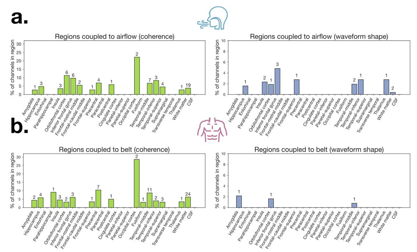
Only a relatively small number of recorded channels passed every test for detailed waveform coupling, particularly when breathing was measured with a belt. The findings also show association rather than cause and effect. Researchers do not yet know whether altering the shape of a breath directly changes the shape of neural activity.
Future experiments could test that question by asking people to deliberately change how they breathe while researchers measure corresponding changes in the brain. Studies could also examine whether breath shape helps predict differences in memory, perception or emotional processing.
“Now that we know that there is this incredibly tight and rich coupling between the shape of each breath and the shape of each brainwave, there’s a whole new world of options that we can explore,” Voytek said.
For now, the findings offer a more detailed view of one of the body’s most familiar rhythms. Breathing is not simply a steady count of inhalations and exhalations. Each breath carries its own timing and shape, and the brain appears to be following along.
These studies explore how respiration interacts with neural activity, memory, perception and large-scale brain networks, providing broader context for the breath-by-breath coupling described in the new research.
Human forebrain neural synchronization and entrainment to breathing during wakefulness, sleep, and external mechanical ventilation: Using intracranial recordings, researchers found widespread synchronization between breathing and human forebrain activity, including when airflow bypassed the nose during mechanical ventilation. The findings suggest that several bodily pathways may carry respiratory information to the brain. (Nature Communications, 2026)
Nasal and oral breathing modes reconfigure brain network dynamics between stabilizing integration and promoting fragmentation: This study used functional MRI to examine how nasal and oral breathing influence changing patterns of communication across brain networks. The results add evidence that the way a person breathes can alter large-scale neural dynamics. (Scientific Reports, 2026)
The respiratory cycle modulates distinct dynamics of affective and perceptual decision-making: Researchers found that inhalation and exhalation influenced the timing and character of decisions involving visual motion and emotional faces. The work provides behavioral evidence that individual phases of breathing can shape perception and affective processing. (PLOS Computational Biology, 2025)
Unmasking the post-expiratory pause: salience network connectivity and its link to psychological factors: This research examined the brief pause that can follow exhalation and connected variations in that respiratory feature with activity in the brain’s salience network and psychological measures. It shows why detailed breath morphology may carry information that breathing rate alone misses. (Cerebral Cortex, 2025)
Single neurons in the thalamus and subthalamic nucleus process cardiac and respiratory signals in humans: Direct recordings from individual human neurons showed that cells in the thalamus and subthalamic nucleus respond to respiratory and cardiac signals. The study provides cellular-level evidence that brain regions beyond the brainstem track internal bodily rhythms. (Proceedings of the National Academy of Sciences, 2024)
Research findings are available online in the journal JNeurosci.
The original story “Every breath you take, every distinct pattern in the brain you make” is published in The Brighter Side of News.
Like these kind of feel good stories? Get The Brighter Side of News’ newsletter.
The post Every breath you take, every distinct pattern in the brain you make appeared first on The Brighter Side of News.


Leave a comment
You must be logged in to post a comment.