Every cell in a developing animal has a history, but ordinary laboratory measurements usually erase that history. A molecular recorder has now helped recover it for more than a million cells in one mouse embryo.
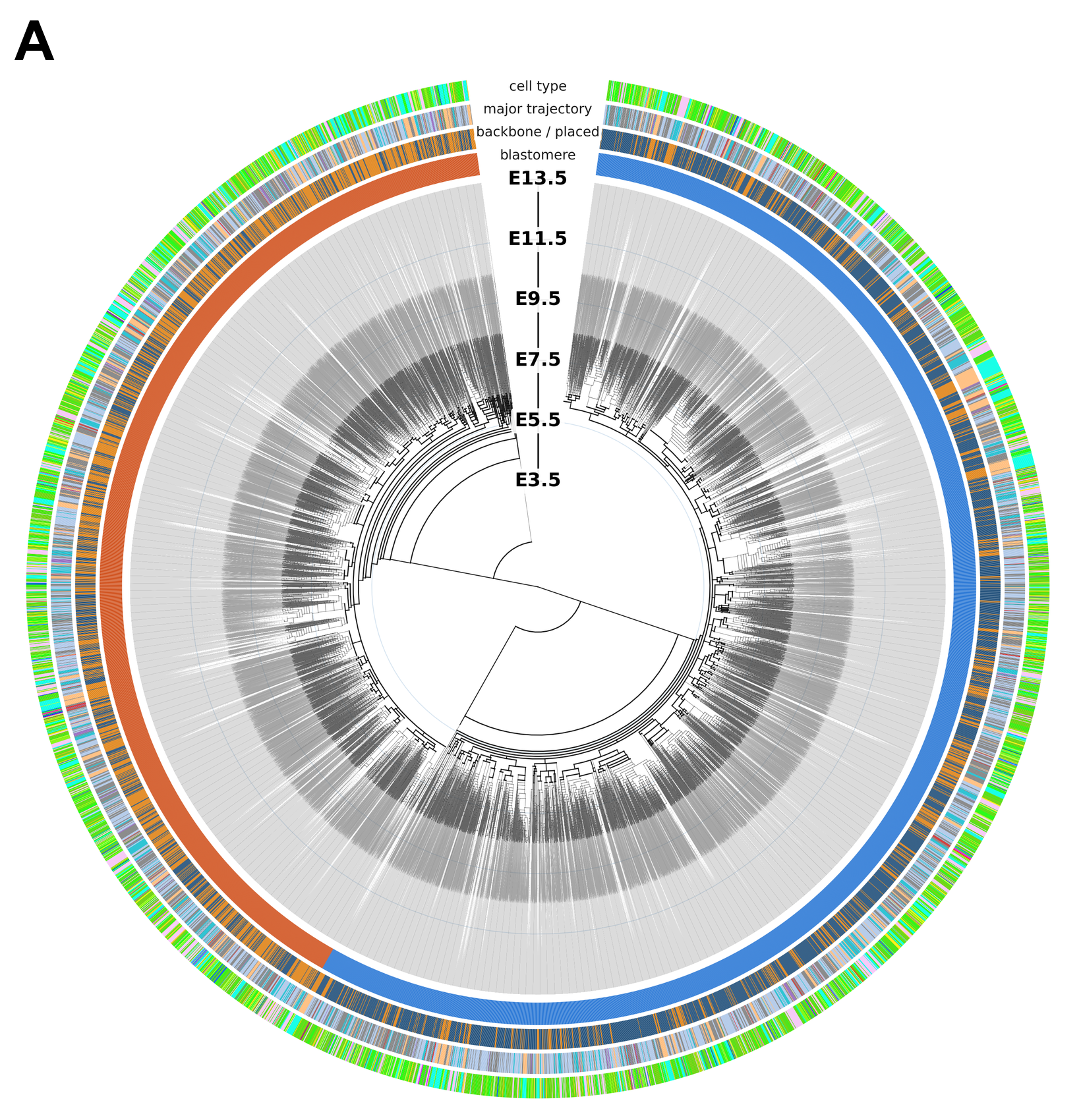
The resulting family tree connects 1,281,141 sampled cells to their ancestry, reaching back to the fertilized egg. It follows development through 13.5 days, when organs have taken shape, and represents about 10% of the embryo’s cells.
Published in Science, the work comes from the Seattle Hub for Synthetic Biology. The partnership brings together the Allen Institute, UW Medicine and Biohub. Jay Shendure, the hub’s scientific director and a UW genome sciences professor, helped lead the study.
The achievement is a large, time-calibrated reconstruction, rather than a complete film of development. It reveals how lineages expand and become associated with particular tissues, while leaving important gaps in the record.
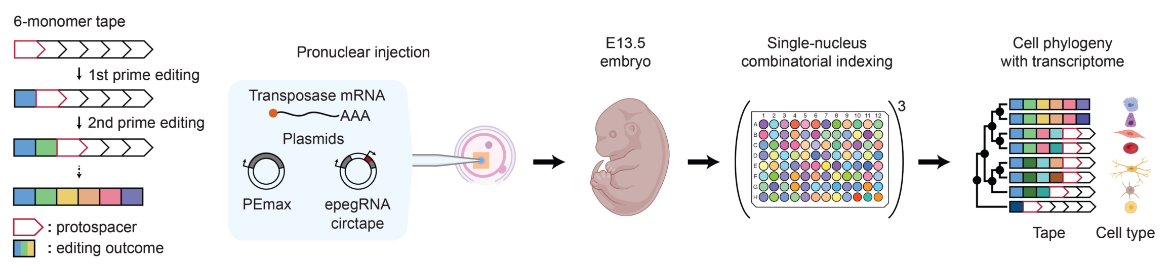
DNA Typewriter uses prime editing to insert short sequences into engineered DNA recording sites, called tapes. Each insertion closes one position and activates the next, preserving the order in which symbols were written.
When a cell divides, its daughters inherit the existing record. Later edits distinguish their descendants, allowing researchers to infer relationships from shared histories. The recorder accumulates marks during development; it does not necessarily write one stamp for every division.
That ordered design helps address problems with earlier recording systems. Methods that cut both DNA strands can delete neighboring records. DNA Typewriter avoids requiring those double-strand breaks, although its records still contain missing data and unexpected edits.
The researchers injected the recording components into fertilized mouse eggs and collected embryos at 13.5 days. One embryo with strong recording activity became the focus of the detailed reconstruction. Its genome contained eleven independently recording tapes.
To retrieve their contents, the team redesigned the tapes to produce circular RNA transcripts. This made the records easier to recover alongside measurements of gene activity.
Scientists profiled nuclei rather than whole cells, helping avoid biases caused by separating mature tissues into individual cells. After quality checks, they obtained gene-expression profiles from 1,584,848 nuclei. Comparing those profiles with an existing mouse-development atlas identified the cells’ types.
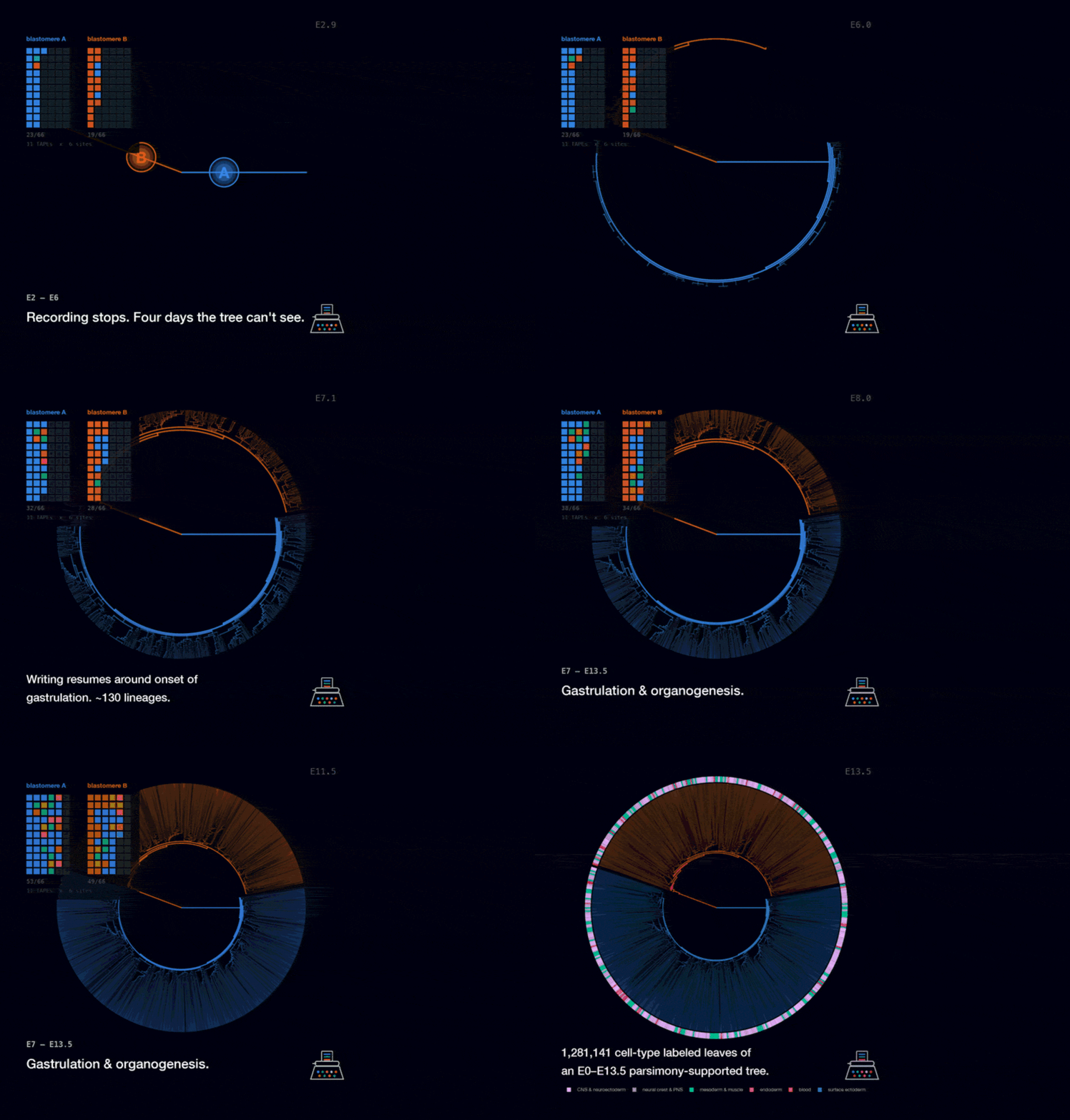
A strong burst of editing near the first division created distinct records in the embryo’s two founding cells. Their descendants could therefore be assigned to two broad branches, which the researchers analyzed separately.
“We were surprised by just how robustly marking happened right at the very first division,” said co-first author Haedong Kim. Those early marks provided an unusually clear starting point for the reconstruction.
Among cells with recovered recording data, about 57.1% were assigned to one founder and 41.9% to the other. Approximately 1% lacked an unambiguous assignment.
Despite that numerical imbalance, the two branches had highly similar cell-type compositions. One contributed more sampled descendants overall, without appearing disproportionately committed to a particular fate.
This finding applies to the embryo itself. Extraembryonic tissues were discarded, so the experiment cannot establish how the founders contributed to those supporting tissues.
Comparing the two branches also helped test whether developmental patterns recurred within the animal. That provides a useful internal check, but it cannot replace repeating the experiment in separate embryos.

The more striking inequality appeared later, around gastrulation, when the embryo establishes its major body layers. Researchers examined 3,849 reconstructed ancestral lineages dated to seven days after fertilization.
Just 170 of those lineages accounted for half the cells in the final tree. A simple model of neutral growth would have required about 720 lineages to supply the same share.
The analysis suggests that unequal contributions developed rapidly and largely plateaued by approximately day nine. It offers a timeline for a phenomenon often studied within individual tissues: some ancestors leave far more descendants than others.
The tree cannot determine whether larger families resulted from faster division, better survival or both. It records surviving lineages, without capturing cells that died along the way.
Spatial position may also matter. By day seven, ancestors occupy different parts of the embryo, where local signals can influence growth and fate. A large contribution therefore does not establish an inherited advantage or a predetermined tissue identity.
Close relatives in the tree often shared a cell type. Across the sampled data, 51% of neighboring sibling pairs had matching identities, about 9.4 times the frequency expected after randomly rearranging labels.
These are tree siblings, not necessarily two daughters of the same observed division. With only a fraction of cells sampled, the nearest available relatives may have missing family members between them.
Broader groups of related cells nevertheless recovered recognizable developmental organization. Their patterns corresponded to blood formation, nervous-system development and other major tissue groupings.
The researchers also used the reference atlas to estimate ancestral gene-expression states. Selected inferred paths resembled established developmental transitions, including the progression toward kidney progenitor cells.
Those ancestral states were computationally estimated, not directly recorded or independently validated. They are exploratory additions to the lineage tree, rather than observations of what every ancestor was doing.
Building the tree required two computational stages. A backbone contained 655,701 cells with more complete records. Another 625,440 were placed near their closest recorded relatives, expanding coverage while adding uncertainty about exact branching positions.
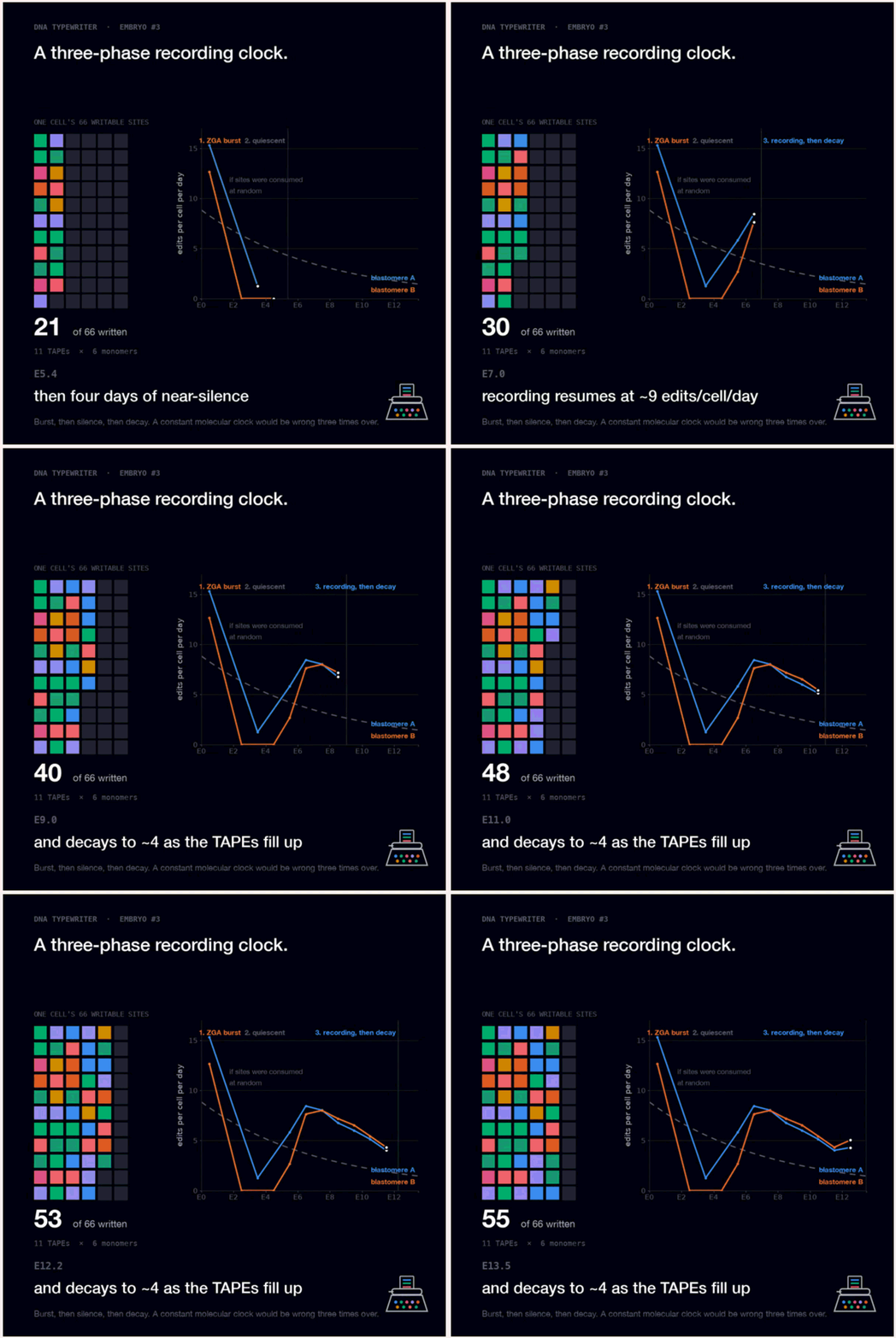
Timing also depends on assumptions. Editing accelerated early, slowed sharply, then recovered around gastrulation. Rates differed between cell types, particularly blood cells, so branch dates are approximations.
The recorder itself may have affected biology. Although broad stress and damage signals were not elevated, some genes near insertion sites changed expression. The researchers cannot yet determine how those changes influenced the tree.
Shendure said the work brings the team closer to “our dream of comprehensively mapping mammalian development.” Such maps could eventually provide references for investigating birth defects, cancer and other conditions in which cellular development changes.
This experiment did not test a disease treatment or establish how a human disorder begins. Its immediate contribution is a resource for studying normal development, with explicit limits on sampling, timing and reproducibility.
The tree is available through NextCell, an interactive browser for exploring cellular ancestry. Independent animals, additional developmental stages and improved recorders will be needed to turn this first large reconstruction into a reliable account of mammalian development.
These resources explain the recording technology and the developmental references that make cellular family trees useful.
A time-resolved, multi-symbol molecular recorder via sequential genome editing: Introduces DNA Typewriter and demonstrates ordered molecular recording in living mammalian cells. (Nature, 2022)
Molecular recording using DNA Typewriter: Describes the experimental workflow for recording the order of molecular events with sequential DNA editing. (Nature Protocols, 2024)
A single-cell time-lapse of mouse prenatal development from gastrula to birth: Provides the gene-expression atlas used to identify cell types and explore inferred ancestral states. (Nature, 2024)
Single-cell, whole-embryo phenotyping of mammalian developmental disorders: Shows how large-scale cellular profiling can reveal developmental changes in genetically altered mouse embryos. (Nature, 2023)
Whole-organism lineage tracing by combinatorial and cumulative genome editing: Demonstrates an earlier genome-editing approach to reconstructing cell ancestry in zebrafish. (Science, 2016)
Research findings are available online in the journal Science.
The original story “Scientists construct a ‘family tree’ containing 1.28 million cells from a single embryo” is published in The Brighter Side of News.
Like these kind of feel good stories? Get The Brighter Side of News’ newsletter.
The post Scientists construct a ‘family tree’ containing 1.28 million cells from a single embryo appeared first on The Brighter Side of News.


Leave a comment
You must be logged in to post a comment.